Review
. 2021 Jan-Feb;15(1):16-26.
doi: 10.1016/j.jcct.2020.11.004. Epub 2020 Nov 13.

Use of cardiac CT amidst the COVID-19 pandemic and beyond: North American perspective

Affiliations
Review

Use of cardiac CT amidst the COVID-19 pandemic and beyond: North American perspective

Vasvi Singh et al. J Cardiovasc Comput Tomogr. 2021 Jan-Feb.

Abstract

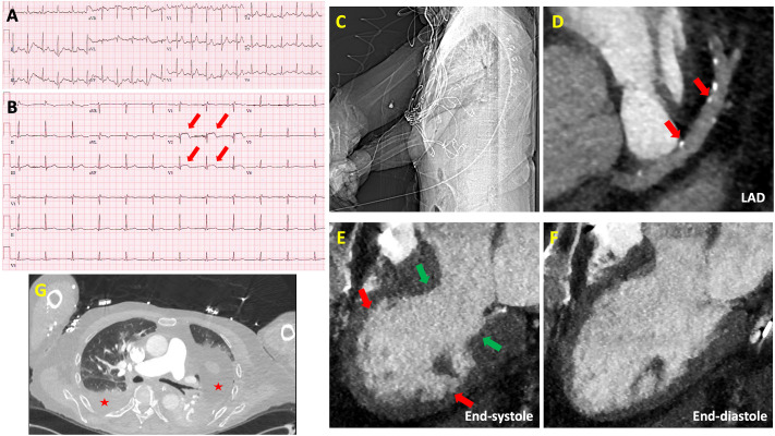
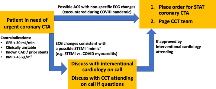
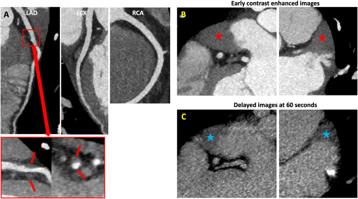
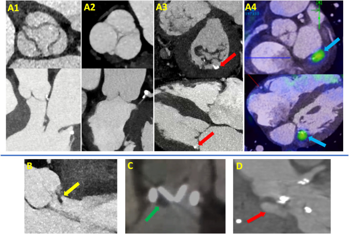
The COVID-19 pandemic has affected patient care deliver throughout the world, resulting in a greater emphasis on efficiently and safety. In this article, we discuss the experiences of several North American centers in utilizing cardiac CT during the pandemic. We also provide a case-based overview which highlights the advantages of cardiac CT in evaluating the following scenarios: (1) patients with possible myocardial injury versus myocardial infarction; (2) patients with acute chest pain; (3) patients with stable chest pain; (4) patients with possible intracardiac thrombus; (5) patients with valvular heart disease. For each scenario, we also provide an overview of various societies recommendations which have highlighted the use of cardiac CT during different phases of the COVID-19 pandemic. We hope that the advantages of cardiac CT that have been realized during the pandemic can help promote wider adoption of this technique and improved coverage and payment by payors.

Keywords: COVID-19; Cardiac CT; Coronary CTA.


Conflict of interest statement

Declaration of competing interest None of the authors of this manuscript have a conflict of interest relevant to this topic.

Figures

Fig. 1
Fig. 1
Role of Coronary CTA in Acute Chest Pain Associated with ST changes. A) Electrocardiogram (ECG) on admission showing sinus tachycardia. B) Subsequent ECG during inpatient admission showing new ST-segment elevations in the anteroseptal leads (red arrows). C) Scout image from cardiac CT showing arms down position during image acquisition. D) Small amount of calcified plaque in the proximal and mid left anterior descending (LAD) artery resulting in minimal (1–24%) stenosis (red arrows). E) 3-chamber end-systolic image of cine-cardiac CT showing akinesis and ballooning of the apical segments, hypokinesis of the mid segments (red arrows) and normal contractility of the basal segments (green arrows) consistent with stress induced cardiomyopathy. F) 3-chamber end-diastolic image of cine-cardiac CT, left ventricular ejection fraction was quantified at 25%. G) Ground-glass opacities and pleural effusions (loculated on the left side) are visualized in bilateral lungs (red stars) consistent with known COVID-19 pneumonia.
Fig. 2
Fig. 2
An institutional experience: Expanded BWH coronary CTA service during COVID-19 pandemic, off-hours (5pm–11pm) coronary CTA workflow.
Fig. 3
Fig. 3
Role of Coronary CTA in Acute Chest Pain in the ED or Observation Unit. A) Normal coronary CTA showing no plaque or stenosis. B1) Coronary CTA showing no plaque or stenosis, B2) Moderate size hiatal hernia (red arrows).
Fig. 4
Fig. 4
Role of Coronary CTA in Stable Chest Pain - To Evaluate for High-risk Coronary Anatomy. A1) 18F-FDG PET/CT scan showing distal esophageal cancer as an FDG avid lesion (blue arrow), A2) Coronary CTA showing a small amount of calcified plaque in the mid LAD causing minimal (1–24%) stenosis (red arrows), and a small amount of calcified and non-calcified plaque in the proximal and mid LCX causing minimal (1–24%) stenosis (green arrows). B1) 4-chamber end-systolic and end-diastolic image of cine-cardiac CT showing hypokinesis of all LV segments, B2) Coronary CTA showing a small amount of calcified plaque in proximal LAD causing minimal (1–24%) stenosis (red arrows), and a small amount of non-calcified plaque in the mid LAD causing mild (25–49%) stenosis (blue arrows).
Fig. 5
Fig. 5
Role of Coronary CTA in Stable Chest Pain – Inconclusive Functional Test. A) Baseline ECG demonstrating left bundle branch block. B) Regadenoson PET myocardial perfusion images showing a dilated left ventricle with a large reversible perfusion defect of moderate intensity involving the entire septum (stress: top row, rest: bottom row). C) Coronary CTA dedicated for bypass graft evaluation showing a patent left internal mammary artery graft to left anterior descending artery, and a patent saphenous venous graft to posterior descending artery.
Fig. 6
Fig. 6
Role of Cardiac CT in Evaluation of Intracardiac Thrombus. A) Coronary CTA showing a small amount of non-calcified plaque in the mid LAD causing mild (25–49%) stenosis (red arrows). B) Early contrast enhanced images showing a filling defect in the left atrial appendage which could represent mixing of contrast due to a low flow state or a thrombus (red stars). C) Delayed images acquired after 60 s showing a persistent filling defect thereby confirming the presence of a left atrial appendage thrombus (blue stars).
Fig. 7
Fig. 7
Role of Cardiac CT in Evaluation of Valvular Heart Disease. A) Cardiac CT showed no valvular vegetations: A1) Aortic valve in short and long axis views, A2) Pulmonic valve in short and long axis views, A3) Mitral and tricuspid valves in short and long axis views, posterior mitral annular calcification (MAC) is seen (red arrows). A4) Fused Cardiac CT images with 18F-FDG PET/CT images demonstrate focal FDG uptake at the mitral annulus at the site of MAC concerning for a nidus of focal infection (blue arrows). B) A large hypodensity (yellow arrow) is seen attached to the aortic valve, most likely representing a vegetation in a patient with gram positive bacteremia. C) A large hypodensity (green arrow) is seen attached to a bileaflet mechanical aortic valve, representing a thrombus in a patient with elevated transvalvular gradient and non-compliance to anticoagulation. D) Aortic root pseudoaneurysm (red arrow) in a patient with recent infective endocarditis.
Fig. 8
Fig. 8
Cardiac CT: One test, many uses.

References

    1. Zoghbi W.A., DiCarli M.F., Blankstein R. Multimodality cardiovascular imaging in the midst of the COVID-19 pandemic: ramping up safely to a new normal. JACC Cardiovasc Imag. 2020;13:1615–1626. - PMC - PubMed
    1. Wood D.A., Mahmud E., Thourani V.H. Safe reintroduction of cardiovascular services during the COVID-19 pandemic: guidance from North American society leadership. J Am Coll Cardiol. 2020;36(7):971–976. - PMC - PubMed
    1. Zoghbi William A., DiCarli Marcelo F., Blankstein Ron, Choi Andrew D., Flachskampf Frank A., Geske Jeffrey B., Grayburn Paul A., Jaffer Farouc A., Kwong Raymond Y., Jonathan A. 2020. Multimodality Cardiovascular Imaging in the Midst of the COVID-19 Pandemic: Ramping up Safely to a New Normal. JACC: Cardiovascular Imaging. - PMC - PubMed
    1. Skulstad H., Cosyns B., Popescu B.A. COVID-19 pandemic and cardiac imaging: EACVI recommendations on precautions, indications, prioritization, and protection for patients and healthcare personnel. Eur Heart J Cardiovasc Imag. 2020;21:592–598. - PMC - PubMed
    1. Skali H., Murthy V.L., Al-Mallah M.H. Guidance and best practices for nuclear cardiology laboratories during the coronavirus disease 2019 (COVID-19) pandemic: an Information Statement from ASNC and SNMMI. J Nucl Med. 2020;27(3):1022–1029. - PMC - PubMed