. 2021 May;157(3):816-833.
doi: 10.1111/jnc.15252. Epub 2020 Dec 17.

Molecular forms of neurogranin in cerebrospinal fluid

Affiliations

Molecular forms of neurogranin in cerebrospinal fluid

Faisal Hayat Nazir et al. J Neurochem. 2021 May.

Abstract

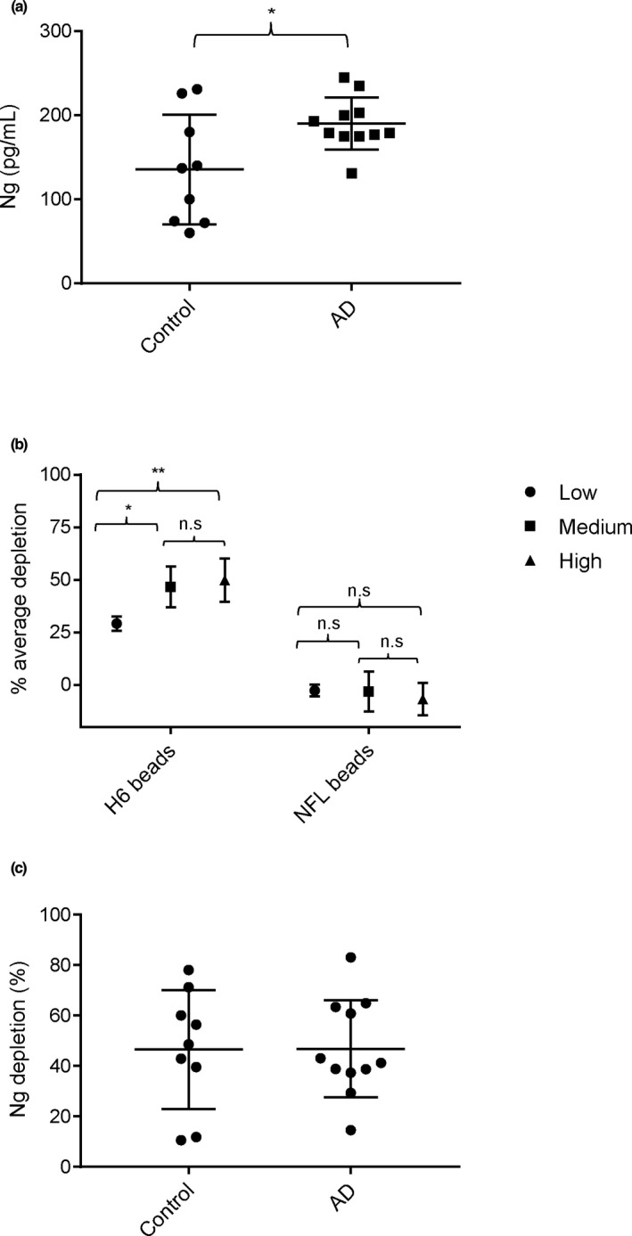
Neurogranin (Ng) is a 78 amino acid neuronal protein and a biomarker candidate for Alzheimer's disease (AD). Ng has been suggested to bind to calmodulin and phosphatidic acid via its centrally located IQ domain. Ng is cleaved within this functionally important domain, yielding the majority of fragments identified in cerebrospinal fluid (CSF), suggesting that cleavage of Ng may be a mechanism to regulate its function. Up to now, Ng has been shown to be present in CSF as both C-terminal fragments as well as full-length protein. To obtain an overview of the different molecular forms of Ng present in CSF, we show by size exclusion chromatography (SEC), immunoblotting, immunoprecipitation, and MS that Ng is present in CSF as several molecular forms. Besides monomeric full-length Ng, also higher molecular weight forms of Ng, and C-terminal- and previously not identified N-terminal fragments were observed. We found by immunodepletion that C-terminal peptides contribute on average to ~50% of the total-Ng ELISA signal in CSF samples. There were no differences in the overall C-terminal fragment/total-Ng ratios between samples from AD and control groups. In addition, we found that monomeric Ng and its C-terminal fragments bind to heparin via a heparin-binding motif, which might be of relevance for their export mechanism from neurons. Taken together, this study highlights the presence of several molecular forms of Ng in CSF, comprising monomeric full-length Ng, and N- and C-terminal truncations of Ng, as well as larger forms of still unknown composition.

Keywords: CSF; heparin-binding motif; neurogranin.


Conflict of interest statement

HZ has served at advisory boards of Denali, Roche Diagnostics, Samumed, CogRx and Wave, has given lectures in symposia sponsored by Fujirebio, Alzecure and Biogen, and is a co‐founder of Brain Biomarker Solutions in Gothenburg AB, a GU Ventures‐based platform company at the University of Gothenburg. KB has served as a consultant or at advisory boards for Alzheon, Abcam, Axon, Biogen, Eli Lilly, Merck, Novartis, Pfizer, and Roche Diagnostics, and is a co‐founder of Brain Biomarker Solutions in Gothenburg AB, a GU Ventures‐based platform company at the University of Gothenburg. The other authors report no conflicts of interest.

Figures

FIGURE 1
FIGURE 1
Ng amino acid (aa) sequence. The aa marked in red text indicates the IQ domain in the Ng sequence. The experimentally estimated epitopes for NG‐H6 and for the NG2 monoclonal antibody and the immunogen for the NG36 monoclonal antibody are indicated (blue arrows)
FIGURE 2
FIGURE 2
Ng is present in CSF as full‐length monomer and as higher molecular weight species. The panels represent immunoblots of pooled CSF concentrates and pooled TBS extracts from human brains (as Ng standard). Human brain extact (“h brain TBS pool”) was used as a positive control for the presence of monomeric Ng. Samples were analyzed on 4%–12% SDS‐PAGE gels at non‐reducing conditions. NG36, NG2 and NG‐H6 (below panels) were the mabs used to detect Ng species. Red circles denote the major bands detected and the arrows indicate their apparent molecular sizes: Ng monomer at approx. 12 kDa, and higher molecular weight species at approx. 35 kDa and 70 kDa. The band at 55 kDa in the NG‐H6 immunoblot most likely represents GAP43, which is known to cross‐react with the NG‐H6 antibody, since that antibody recognizes an epitope in the IQ domain which is present both in Ng and GAP43 (see also Figure S1). WB ctrl, western blot control probed with mouse IgGκ‐binding protein‐HRP only
FIGURE 3
FIGURE 3
CSF Ng is present in various fractions after size exclusion chromatography (SEC). Pooled CSF was concentrated by ultrafiltration, followed by SEC. The elution positions of SEC size markers (in kDa) are indicated by arrows below the graph. **, major peak at ~25 kDa, which is close to the reported size range (~22 kDa) of monomeric Ng in Triton X‐100 containing buffer (Kumar et al., 2013). * and ***, minor peaks, before and after the main peak containing higher and lower molecular weight species, respectively
FIGURE 4
FIGURE 4
Neurogranin is present in different molecular forms in CSF. Size exclusion chromatography (SEC) fractions (fractions 14–43; on adjacent blots) were collected and processed for SDS‐PAGE at non‐reducing conditions followed by immunoblotting (mab NG36). The positions of SDS‐PAGE size markers are shown on the left side and the fraction numbers are shown above each lane on the blots. The SEC column was initially calibrated with SEC size markers; their elution positions are indicated by arrows below the blots. Ng_Adx, synthetic Ng1‐78 (as positive control). This lane is shown here at lower exposure than the rest of the blot to avoid overexposure of the bands. Fraction “bef. SEC” represents the concentrated CSF sample before SEC, but 2.5x less volume of this sample was used for blotting, as compared to the samples from the column fractions. The rectangle shown in green corresponds to molecular weight species of Ng above 12 kDa, the rectangle shown in blue corresponds to ~12 kDa and the rectangle shown in red corresponds to Ng species of ~6 kDa
FIGURE 5
FIGURE 5
Neurogranin peptides identified by MS/MS in immunoprecipitates. (a) A pool of CSF was concentrated by ultrafiltration and then size‐separated on a SEC column. Ng peptides were immunoprecipitated from the concentrated CSF pool before (blue bars) and after SEC separation (red bars) by a mixture of bead‐immobilized mabs NG36 and NG‐H6. (b) Neurogranin peptides identified in the non‐concentrated CSF pool [same pool as in (a)] by NG36, NG‐H6, and a mixture of NG36 and NG‐H6. Only peptides which had at least one peak area (among the three immunoprecipitations) of more than 1% of the largest peak area are shown. The heights of the bars in (a) and (b) reflect peak areas of identified peptides (sum of all major known modifications for each peptide)
FIGURE 6
FIGURE 6
Schematic representation of the workflow to analyze the proportion of C‐terminal Ng fragments to “total‐Ng”. Total‐Ng comprises all Ng species detectable by the C‐terminal sandwich ELISA used (= C‐terminal fragments and full‐length Ng). Individual CSF samples were either immunodepleted of N‐terminal and full‐length Ng using NG‐H6 beads, or using NFL‐21 control beads, followed by analysis of the supernatants using an in‐house sandwich ELISA with NG36 as capture antibody and NG2 as the detection antibody
FIGURE 7
FIGURE 7
Immunodepletion of full‐length Ng and Ng fragments (starting N‐terminally of ~aa 43) from CSF using NG‐H6 beads. (a) Levels of Ng in control and AD samples before immunodepletion by sandwich ELISA; p = .04 (unpaired t‐test with Welsh's correction); N, number of individuals: control = 9 and AD = 11; median control = 137, median AD = 179; control SD = 65.3, AD SD = 31.1. The mean and standard error of mean (SEM) are indicated in the scatter plot. (b) Depletion levels reach saturated maximum levels. NG‐H6 and NFL‐21 (control) mab were cross‐linked separately to beads. Those beads were then diluted with PBS to three concentrations of beads (high, medium, low; five‐fold dilution steps) and used at equal bead suspension volumes to deplete Ng species from CSF. Average depletion (all samples) reached a plateau at middle and high bead concentrations of NG‐H6 beads, but not at low bead concentration. NFL control beads did not show depletion. One‐way ANOVA followed by Tukey's post‐hoc analysis shows the significant Ng reduction when compared between low and medium NG‐H6 antibody bead concentration and between low and high NG‐H6 bead concentration. No significant (n.s) differences were observed between medium and high NG‐H6 bead concentration; the immunodepletion had reached saturation at these concentrations. No depletion of Ng was detected with NFL‐21 antibody at any NFL‐21 bead concentration (high, medium, and low) and therefore no significant differences were found using one‐way ANOVA followed by Tukey's post‐hoc analysis. The data points represent the percentage of Ng depleted (0% depletion = no beads but PBS added); the error bars indicate SEM, n = 20 for each concentration of beads, p < .05 *, p < .01**. NG‐H6 mab depletes Ng significantly (unpaired t‐test) at all bead concentrations used, as compared to NFL‐21 antibody (NG‐H6 low versus NFL‐21 low, p < .001; NG‐H6 medium versus NFL‐21 medium, p < .001; NG‐H6 high versus NFL‐21 high, p < .001. (c) Ng depletion (%) in control and AD samples after immunodepletion with NG‐H6 beads using medium bead concentration; p = .97 (unpaired t‐test with Welsh's correction); N, number of individuals: control = 9 and AD = 11; median control = 48.5, median AD = 41.2; control SD = 23.58 and AD SD = 19.3. The mean and standard error of mean (SEM) are indicated in the scatter plot
FIGURE 8
FIGURE 8
(a) Full‐length Ng and C‐terminal Ng peptides containing KKIK aa sequence motif, but not N‐terminal peptides NgX…42 ("X" denoting undefined amino acid in sequence), bind to heparin beads. Synthetic and recombinant (rec.) Ng and Ng peptides, either exposed to PBS control (“‐”, in stained gel) or heparin beads (“+”), were separated under reducing conditions on a 4%–12% SDS PAGE gel. The gel was then sequentially stained by silver stain and Coomassie stain. The red circles point to the disappearance of the peptide bands because of binding of the peptides to heparin beads. MW, molecular weight markers (kDa). (b) Summary of the heparin‐binding properties of full‐length Ng and Ng peptides

References

    1. Alvarez‐Castelao, B., & Schuman, E. M. (2015). The regulation of synaptic protein turnover. The Journal of Biological Chemistry, 290, 28623–28630. 10.1074/jbc.R115.657130 - DOI - PMC - PubMed
    1. Alzoubi, K. H., Gerges, N. Z., & Alkadhi, K. A. (2005). Levothyroxin restores hypothyroidism‐induced impairment of LTP of hippocampal CA1: electrophysiological and molecular studies. Experimental Neurology, 195(2), 330–341. - PubMed
    1. Bahler, M., & Rhoads, A. (2002). Calmodulin signaling via the IQ motif. FEBS Letters, 513, 107–113. 10.1016/S0014-5793(01)03239-2 - DOI - PubMed
    1. Baudier, J., Deloulme, J. C., Alain Van Dorsselaer, A. V., Black, D., & Matthes, H. W. D. (1991). Purification and Characterization of a brain specific Protein Kinase C Substrate, Neurogranin (p 17). The Journal of Biological Chemistry, 266, 229–237. - PubMed
    1. Becker, B., Nazir, F. H., Brinkmalm, G., Camporesi, E., Kvartsberg, H., Portelius, E., Boström, M., Kalm, M., Höglund, K., Olsson, M., Zetterberg, H., & Blennow, K. (2018). Alzheimer‐associated cerebrospinal fluid fragments of neurogranin are generated by Calpain‐1 and prolyl endopeptidase. Molecular Neurodegeneration, 13, 47. 10.1186/s13024-018-0279-z - DOI - PMC - PubMed

Publication types

Associated data