. 2021 Apr;36(4):739-756.
doi: 10.1002/jbmr.4223. Epub 2020 Dec 18.

Impact of Genetic and Pharmacologic Inhibition of Myostatin in a Murine Model of Osteogenesis Imperfecta

Affiliations

Impact of Genetic and Pharmacologic Inhibition of Myostatin in a Murine Model of Osteogenesis Imperfecta

Catherine L Omosule et al. J Bone Miner Res. 2021 Apr.

Abstract

Osteogenesis imperfecta (OI) is a genetic connective tissue disorder characterized by compromised skeletal integrity, altered microarchitecture, and bone fragility. Current OI treatment strategies focus on bone antiresorptives and surgical intervention with limited effectiveness, and thus identifying alternative therapeutic options remains critical. Muscle is an important stimulus for bone formation. Myostatin, a TGF-β superfamily myokine, acts through ActRIIB to negatively regulate muscle growth. Recent studies demonstrated the potential benefit of myostatin inhibition with the soluble ActRIIB fusion protein on skeletal properties, although various OI mouse models exhibited variable skeletal responses. The genetic and clinical heterogeneity associated with OI, the lack of specificity of the ActRIIB decoy molecule for myostatin alone, and adverse events in human clinical trials further the need to clarify myostatin's therapeutic potential and role in skeletal integrity. In this study, we determined musculoskeletal outcomes of genetic myostatin deficiency and postnatal pharmacological myostatin inhibition by a monoclonal anti-myostatin antibody (Regn647) in the G610C mouse, a model of mild-moderate type I/IV human OI. In the postnatal study, 5-week-old wild-type and +/G610C male and female littermates were treated with Regn647 or a control antibody for 11 weeks or for 7 weeks followed by a 4-week treatment holiday. Inhibition of myostatin, whether genetically or pharmacologically, increased muscle mass regardless of OI genotype, although to varying degrees. Genetic myostatin deficiency increased hindlimb muscle weights by 6.9% to 34.4%, whereas pharmacological inhibition increased them by 13.5% to 29.6%. Female +/mstn +/G610C (Dbl.Het) mice tended to have similar trabecular and cortical bone parameters as Wt showing reversal of +/G610C characteristics but with minimal effect of +/mstn occurring in male mice. Pharmacologic myostatin inhibition failed to improve skeletal bone properties of male or female +/G610C mice, although skeletal microarchitectural and biomechanical improvements were observed in male wild-type mice. Four-week treatment holiday did not alter skeletal outcomes. © 2020 American Society for Bone and Mineral Research (ASBMR).

Keywords: BONE-MUSCLE INTERACTIONS; COL1A2; OSTEOGENESIS IMPERFECTA (OI); PRECLINICAL STUDIES; TGF-Β.


Conflict of interest statement

Disclosures

SK provided the Regn647 and Regn1945 (Regeneron Pharmaceuticals Inc., Tarrytown, NY, USA), is an employee of Regeneron Pharmaceuticals, and owns stock of this company. LM, JM, and AR are employees of Regeneron Pharmaceuticals. All other authors state that they have no conflicts of interest.

Figures

Fig 1.
Fig 1.
Verification of myostatin deficiency in genetic model. (A) Schematic of congenital +/mstn and +/G610C breeding strategy with the resultant offspring genotypes. (B) Circulating serum myostatin (GDF8, pg/mL) levels in 16-week-old male (solid data points) and female (open data points), wild-type (Wt, black circle), heterozygote myostatin deficient (+/mstn, magenta square), heterozygote G610C (+/G610C, orange diamond), and double heterozygote +/mstn and +/G610C (Dbl.Het, green triangle) offspring (n = 8–15). Data were analyzed by MANOVA and AR (1) model. No genotype by sex or by dam genotype interactions were found. Offspring genotype main effect was evaluated. Values are median with interquartile range. The p values ≤ 0.1 are indicated and considered significant.
Fig 2.
Fig 2.
Congenital myostatin deficiency increases body weight in the +/G610C mouse. (A) Body weights (g) of male (solid data points) and female (open data points) wild-type (Wt, black circle), heterozygote myostatin deficient (+/mstn, magenta square), heterozygote G610C (+/G610C, orange diamond), and double heterozygote +/mstn and +/G610C (Dbl.Het, green triangle) at 16 weeks of age (n = 12–34). (B) Weekly weights of male Wt, +/mstn, +/G610C, and Dbl. Het offspring starting from 3 weeks to 16 weeks of age (n = 11–28). (C) Weekly weights of female Wt, +/mstn, +/G610C, and Dbl. Het offspring starting from 3 weeks to 16 weeks of age (n = 11–37). Data were analyzed by MANOVA and AR (1) model. No genotype by sex or by dam genotype interactions were found. Offspring genotype main effect was evaluated. Values are median with interquartile range. The p values ≤ 0.1 are indicated and considered significant for +/G610C versus Dbl.Het comparisons (table; bottom).
Fig 3.
Fig 3.
Inherent myostatin deficiency increases wet muscle weights (mg) of the (A) gastrocnemius, (B) quadriceps, (C) tibialis anterior, and (D) plantaris at 16 weeks of age in wild-type (Wt, black circle), heterozygote myostatin deficient (+/mstn, magenta square), heterozygote G610C (+/G610C, orange diamond), and double heterozygote +/mstn and +/G610C (Dbl.Het, green triangle) offspring (n = 11–34). Data were analyzed by MANOVA. No genotype by sex or by dam genotype interactions were found. Offspring genotype main effect was evaluated. Values are median with interquartile range. The p values ≤ 0.1 are indicated and considered significant.
Fig 4.
Fig 4.
Inherent myostatin deficiency has minimal impact on femoral bone microarchitecture in +/G610C mice. μCT analyses of 16-week-old male and female wild-type (Wt, black circle), heterozygote myostatin deficient (+/mstn, magenta square), heterozygote G610C (+/G610C, orange diamond), and double heterozygote +/mstn and +/G610C (Dbl.Het, green triangle) offspring femora. Trabecular bone parameters: (A) total volume (mm3), (B) bone volume (mm3), (C) trabecular spacing (mm), (D) trabecular bone volume fraction (BV/TV), (E) trabecular number (Tb.N; 1/mm), (F) trabecular thickness (Tb.Th; mm), and (G) bone mineral density (BMD; g/cm3). Data were analyzed by MANOVA. No genotype by sex or by dam genotype interactions were found. Offspring genotype main effect was evaluated. Values are median with interquartile range. The p values ≤ 0.1 are indicated and considered significant; n = 8–16.
Fig 5.
Fig 5.
Inherent myostatin deficiency failed to improve femoral cortical architecture in +/G610C mice. μCT analyses of 16-week-old male and female wild-type (Wt, black circle), heterozygote myostatin deficient (+/mstn, magenta square), heterozygote G610C (+/G610C, orange diamond), and double heterozygote +/mstn and +/G610C (Dbl.Het, green triangle) offspring mid-diaphyseal femoral cortical bone. Cortical bone parameters: (A) bone area (mm2), (B) total area (mm2), (C) bone area fraction (bone area/total area; BV/TV), and (D) polar moment of inertia (pMOI; mm4). Data were analyzed by MANOVA. No genotype by sex or by dam genotype interactions were found. Offspring genotype main effect was evaluated. Values are median with interquartile range. The p values ≤ 0.1 are indicated and considered significant; n = 7–17.
Fig 6.
Fig 6.
Inherent myostatin deficiency does not improve femoral bone strength in +/G610C mice. Three-point bend biomechanical testing of 16-week-old male and female (Wt, black circle), heterozygote myostatin deficient (+/mstn, magenta square), heterozygote G610C (+/G610C, orange diamond), and double heterozygote +/mstn and +/G610C (Dbl.Het, green triangle) offspring femurs. Three-point bend analysis was performed (A). Biomechanical parameters: (B) maximum load (N), (C) yield load (N), (D) post-yield displacement (mm), (E) stiffness (N/mm), and (F) work to fracture (Nmm). Data were analyzed by MANOVA. No genotype by sex or by dam genotype interactions were found. Offspring genotype main effect was evaluated. Values are median with interquartile range. The p values ≤ 0.1 are indicated and considered significant; n = 6–17.
Fig 7.
Fig 7.
Anti-myostatin antibody treatment increases body weights of Wt and +/G610C mice after only 1 week of treatment. (A) Schematic of postnatal myostatin inhibition treatment regimen; Wt and +/G610C mice were treated twice weekly with either a monoclonal anti-myostatin antibody (Regn647) for 11 weeks (Mstn-ab 11wk, red) or for 7 weeks followed by a 4-week treatment hiatus (Mstn-ab 7wk, blue) or control antibody for 11 weeks (Regn1945, ctrl-ab, black). (B) Body weights of male and female Wt and +/G610C mice at 16 weeks of age. (C, D) Weekly body weights of male and female mice starting at 5 weeks of age, respectively. Control treatment (black symbols) is administered from 5 to 16 weeks of age. Mstn-ab treatment is administered from 5 to 11 weeks (green symbols) at which point the cohort is randomly divided into two groups: Mstn-ab TRT holiday (blue; no further Regn647 treatment [“treatment holiday”] until euthanization at 16 weeks of age) and Mstn-ab Wks 11–16 (red; continuous Mstn-ab treatment until euthanization at 16 weeks of age). For significance values, see Tables 2 and 3. Data were analyzed by MANOVA (B) and AR (1) model (C, D). No significant genotype by sex by treatment interaction was found. (B) Body weight values are median with interquartile range. The p values ≤ 0.1 are indicated and considered significant. (C, D) Values are means. n = 9–12.
Fig 8.
Fig 8.
Postnatal myostatin inhibition increases hindlimb muscle mass in Wt and +/G610C mice. Male and female Wt and +/G610C mice were treated twice weekly with either a monoclonal anti-myostatin antibody (Regn647) for 11 weeks (Mstn-ab Wks 11–16, red) or for 7 weeks followed by a 4-week treatment hiatus (Mstn-ab TRT holiday, blue) or a control antibody for 11 weeks (Regn1945, ctrl-ab, black). Weights of (A) gastrocnemius, (B) quadriceps, (C) tibialis anterior, and (D) plantaris muscles at 16 weeks of age. Data were analyzed by MANOVA. Values are median with interquartile range. The p values ≤ 0.1 are indicated and considered significant. n = 9–12.
Fig 9.
Fig 9.
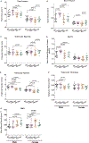
Postnatal myostatin inhibition improves femoral trabecular bone parameters of Wt male mice. Femoral μCT analyses of 16-week-old male and female Wt and +/G610C mice treated twice weekly with either a monoclonal anti-myostatin antibody (Regn647) for 11 weeks (Mstn-ab Wks 11–16, red) or for 7 weeks followed by a 4-week treatment hiatus (Mstn-ab TRT holiday, blue) or a control antibody for 11 weeks (Regn1945, ctrl-ab, black). Trabecular bone parameters: (A) total volume (mm3), (B) bone volume (mm3), (C) trabecular spacing (mm), (D) trabecular bone volume fraction (BV/TV), (E) trabecular number (Tb.N; 1/mm), (F) trabecular thickness (Tb.Th; mm), and (G) bone mineral density (BMD; g/cm3). Data were analyzed by MANOVA. Values are median with interquartile range. The p values ≤ 0.1 are indicated and considered significant. n = 6–11.
Fig 10.
Fig 10.
Mid-diaphyseal femoral cortical bone parameters are improved in Wt males by postnatal myostatin inhibition. μCT analyses of mid-diaphyseal cortical bone of 16-week-old male and female Wt and +/G610C mice treated twice weekly with either a monoclonal anti-myostatin antibody (Regn647) for 11 weeks (Mstn-ab Wks 11–16, red) or for 7 weeks followed by a 4-week treatment hiatus (Mstn-ab TRT holiday, blue) or a control antibody for 11 weeks (Regn1945, ctrl-ab, black). Cortical bone parameters: (A) bone area (mm2), (B) total area (mm2), (C) bone area fraction (BA/TA), and (D) polar moment of inertia (mm4). Data were analyzed by MANOVA. Values are median with interquartile range. The p values ≤ 0.1 are indicated and considered significant. n = 6–11.
Fig 11.
Fig 11.
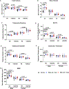
Postnatal myostatin inhibition improves biomechanical properties of male Wt mouse femurs. Three-point bend biomechanical testing of femurs from 16-week-old male and female Wt and +/G610C mice treated twice weekly with either a monoclonal anti-myostatin antibody (Regn647) for 11 weeks (Mstn-ab Wks 11–16, red) or for 7 weeks followed by a 4-week treatment hiatus (Mstn-ab TRT holiday, blue) or a control antibody for 11 weeks (Regn1945, ctrl-ab, black). Biomechanical parameters: (A) maximum load (N), (B) yield load (N), (C) post-yield displacement (mm), (D) stiffness (N/mm), and (E) work to fracture (Nmm). Data were analyzed by MANOVA. Values are median with interquartile range. The p values ≤ 0.1 are indicated and considered significant. n = 6–11.

References

    1. Forlino A, Marini JC. Osteogenesis imperfecta. Lancet. 2016;387 (10028):1657–71. - PMC - PubMed
    1. Morello R Osteogenesis imperfecta and therapeutics. Matrix Biol. 2018;71–72:294–312. - PMC - PubMed
    1. Folkestad L, Hald JD, Hansen S, et al. Bone geometry, density, and microarchitecture in the distal radius and tibia in adults with osteogenesis imperfecta type I assessed by high-resolution pQCT. J Bone Miner Res. 2012;27(6):1405–12. - PubMed
    1. Patel RM, Nagamani SCS, Cuthbertson D, et al. A cross-sectional multicenter study of osteogenesis imperfecta in North America—results from the linked clinical research centers. Clin Genet. 2015;87(2): 133–40. - PMC - PubMed
    1. Oestreich AK, Carleton SM, Yao X, et al. Myostatin deficiency partially rescues the bone phenotype of osteogenesis imperfecta model mice. Osteoporos Int. 2016;27(1):161–70. - PMC - PubMed

Publication types