. 2021 Jul;21(7):2360-2371.
doi: 10.1111/ajt.16417. Epub 2021 Jan 4.

Interleukin 6 trans-signaling is a critical driver of lung allograft fibrosis

Affiliations

Interleukin 6 trans-signaling is a critical driver of lung allograft fibrosis

David S Wheeler et al. Am J Transplant. 2021 Jul.

Abstract

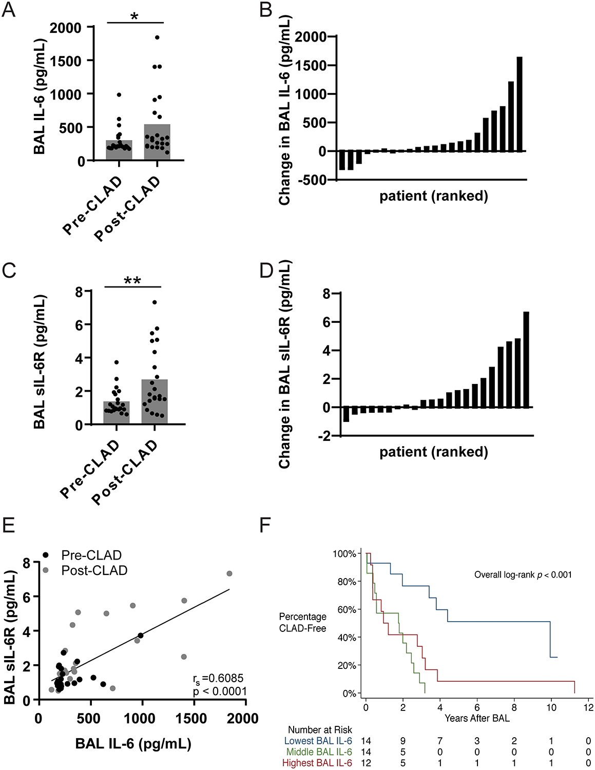
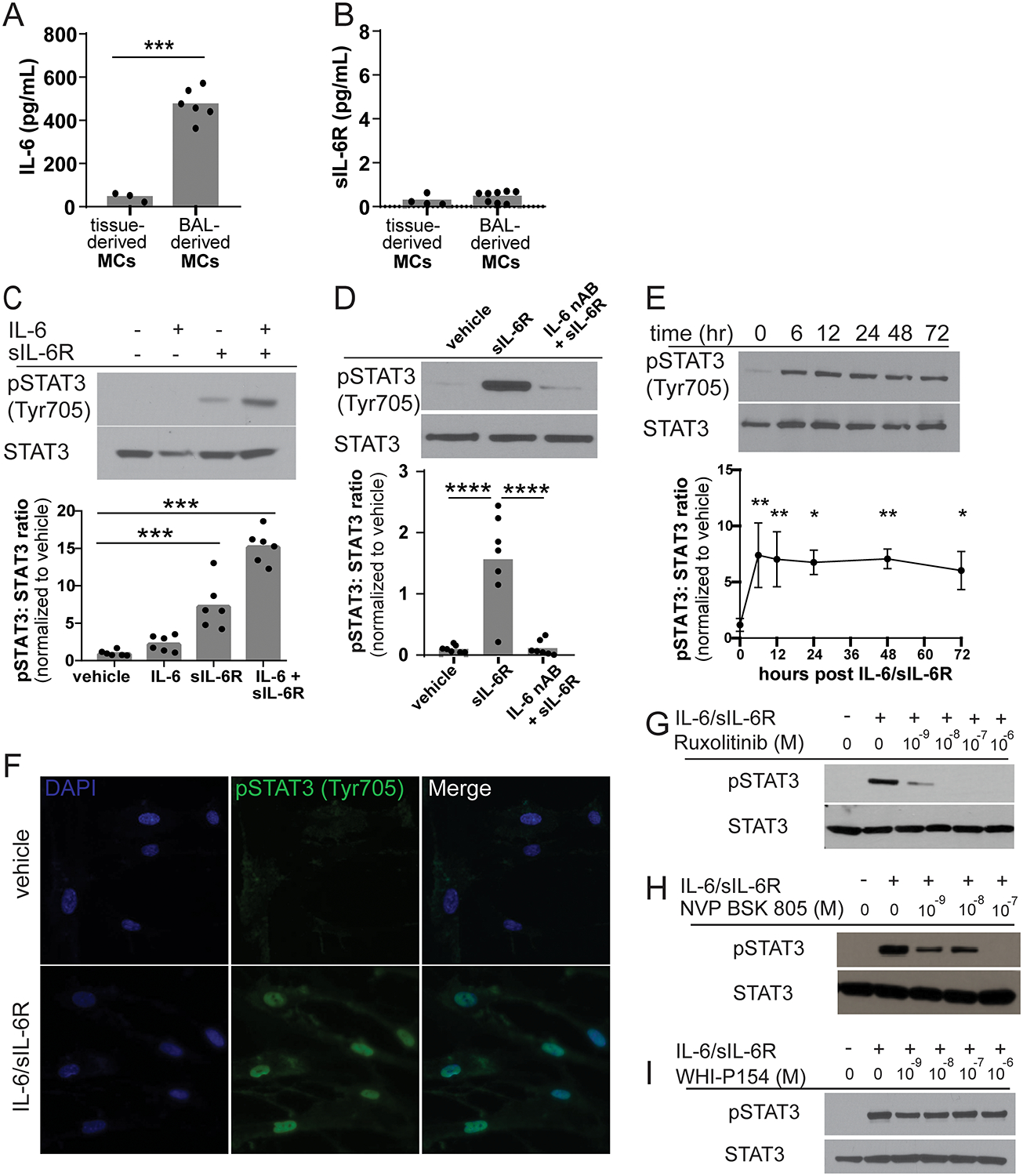
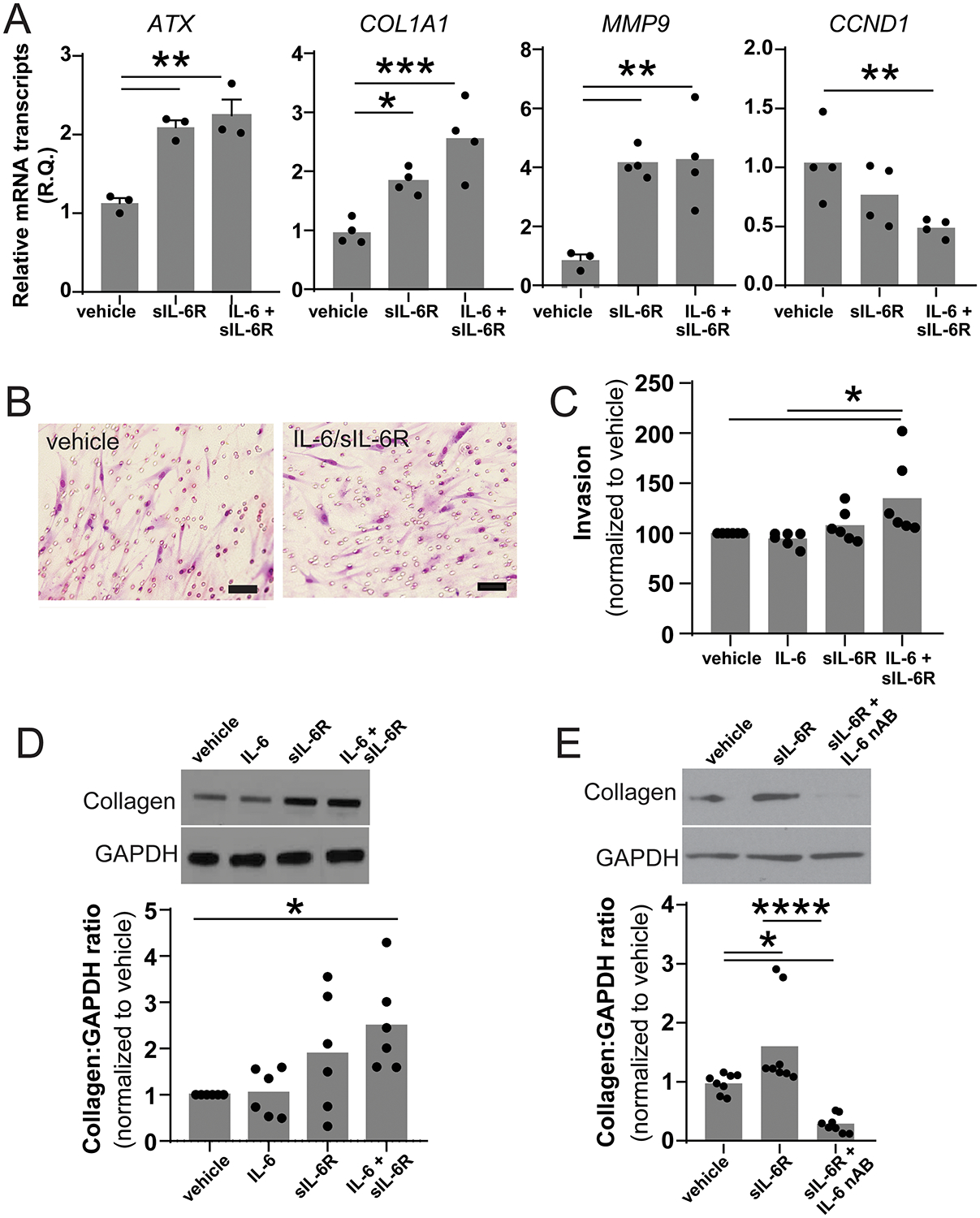
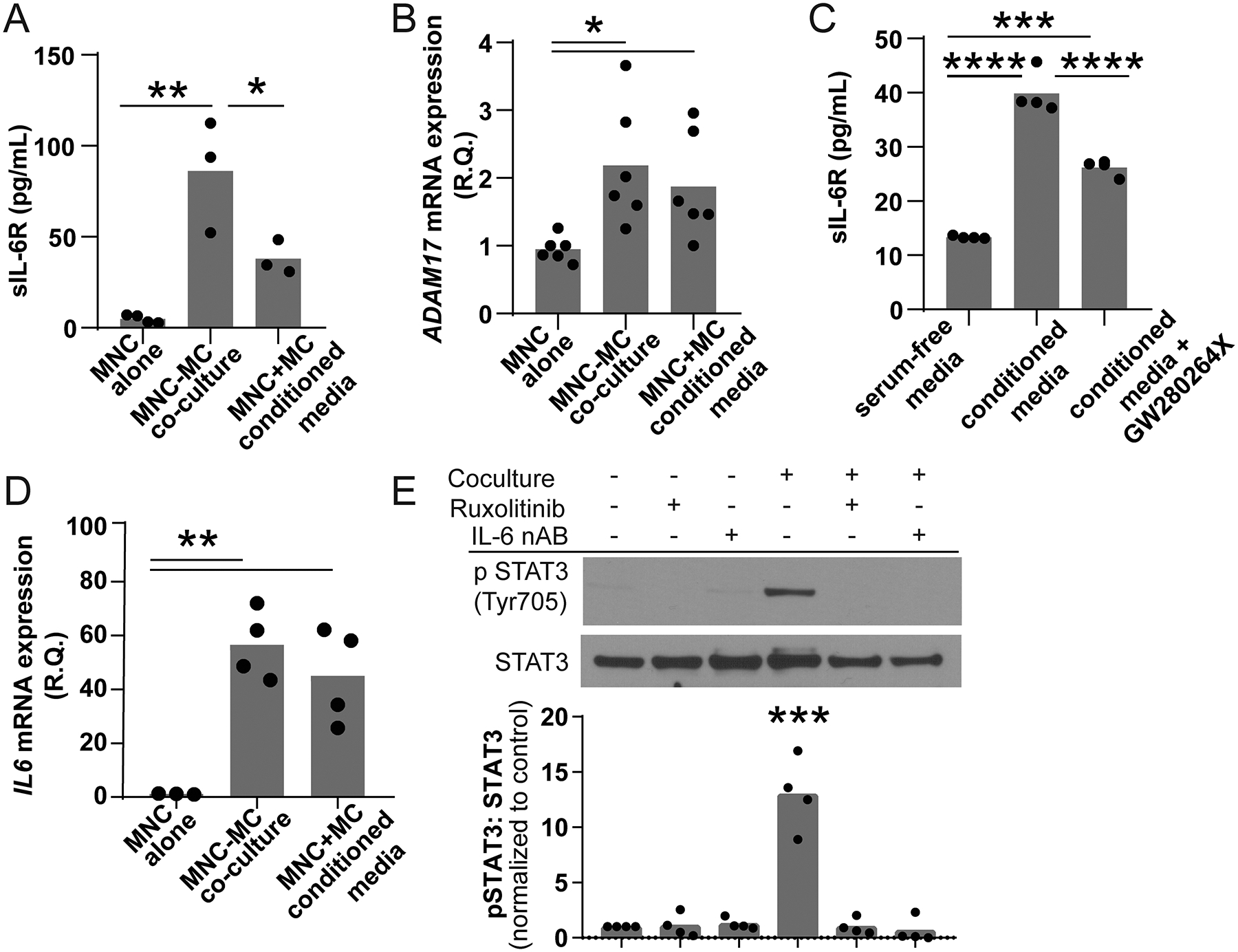
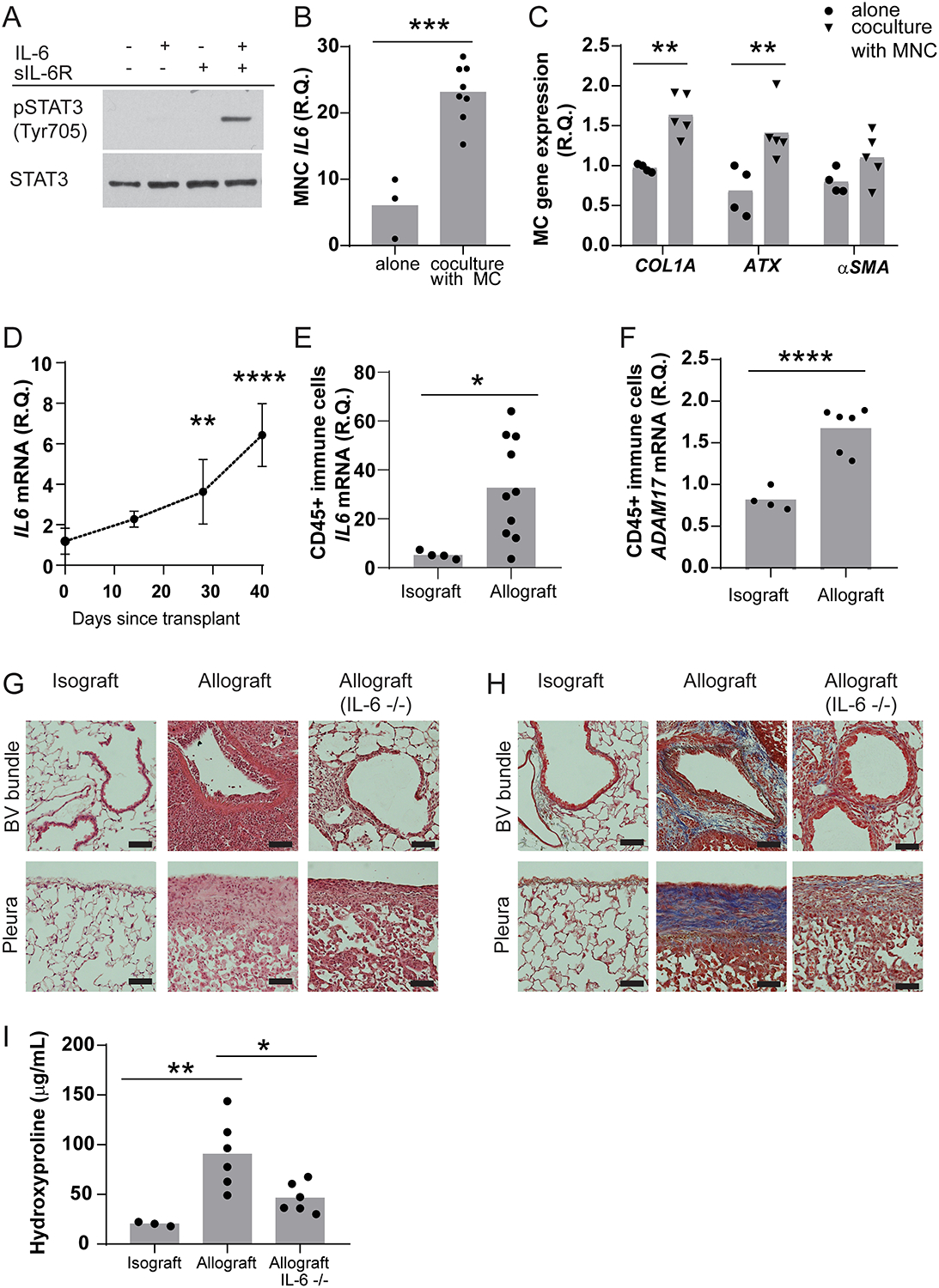
Histopathologic examination of lungs afflicted by chronic lung allograft dysfunction (CLAD) consistently shows both mononuclear cell (MNC) inflammation and mesenchymal cell (MC) fibroproliferation. We hypothesize that interleukin 6 (IL-6) trans-signaling may be a critical mediator of MNC-MC crosstalk and necessary for the pathogenesis of CLAD. Bronchoalveolar lavage (BAL) fluid obtained after the diagnosis of CLAD has approximately twofold higher IL-6 and soluble IL-6 receptor (sIL-6R) levels compared to matched pre-CLAD samples. Human BAL-derived MCs do not respond to treatment with IL-6 alone but have rapid and prolonged JAK2-mediated STAT3 Tyr705 phosphorylation when exposed to the combination of IL-6 and sIL-6R. STAT3 phosphorylation within MCs upregulates numerous genes causing increased invasion and fibrotic differentiation. MNC, a key source of both IL-6 and sIL-6R, produce minimal amounts of these proteins at baseline but significantly upregulate production when cocultured with MCs. Finally, the use of an IL-6 deficient recipient in a murine orthotopic transplant model of CLAD reduces allograft fibrosis by over 50%. Taken together these results support a mechanism where infiltrating MNCs are stimulated by resident MCs to release large quantities of IL-6 and sIL-6R which then feedback onto the MCs to increase invasion and fibrotic differentiation.

Keywords: animal models: murine; basic (laboratory) research / science; bronchiolitis obliterans (BOS); cellular biology; cytokines / cytokine receptors; fibrosis; lung transplantation / pulmonology; translational research / science.


Conflict of interest statement

DISCLOSURE

The authors of this manuscript have no conflicts of interest to disclose as described by the American Journal of Transplantation.

Figures

Figure 1.
Figure 1.. BAL fluid obtained from patients with CLAD demonstrate elevated levels of IL-6 and sIL-6R when compared to CLAD free controls.
(A) BAL concentrations of IL-6 were measured by ELISA for 22 matched pre-CLAD and post-CLAD samples and were compared using a matched pair Wilcoxon test. Post-CLAD samples had significantly higher IL-6 levels (535.2±476.7pg/mL) compared to CLAD free controls (296.0±193.2pg/mL, Wilcoxon test p=0.0251). (B) Absolute change in BAL IL-6 concentration per patient ranked in ascending order. (C) Post-CLAD BAL samples also had higher levels of sIL-6R (2.670±1.96pg/mL), measured by ELISA, compared to CLAD free controls (1.385±0.77pg/mL; Wilcoxon test p=0.0074). (D) Absolute change in BAL sIL-6R concentration per patient ranked in ascending order. (E) There is a significant linear correlation between IL-6 and sIL-6R levels in both pre-CLAD (Spearman’s correlation=0.3056, p=0.0076), post-CLAD (Spearman’s correlation=0.5011, p=0.0002), and combined groups (shown). (F) The parent cohort of 40 one-year surveillance bronchoscopies was subdivided into tertiles based on BAL IL-6 levels (<180 pg/mL, 180–212 pg/mL, >212 pg/mL) and compared using standard Kaplan-Meyer analysis. Patients in the lowest tertile had significantly longer time to CLAD onset compared to patients in the middle or upper tertiles (media survival 4.402 vs. 1.777 vs. 1.077 years respectively, p=0.001).
Figure 2.
Figure 2.. IL-6 trans-signaling stimulates prolonged STAT3 phosphorylation via a JAK2-dependent mechanism in human BAL-derived MCs.
(A) Confluent cultures of pulmonary tissue-derived fibroblasts and BAL-derived MC were grown in serum free media for 24 hours. The media was removed and IL-6 and sIL-6R concentrations were measured by ELISA. Tissue-derived fibroblasts secreted approximately one-tenth of the IL-6 (44.75±20.49pg/mL, n=3) produced by BAL-derived MCs cells (474.5±74.11pg/mL, n=6, p<0.0001). (B) Neither tissue-derived fibroblasts or BAL-derived MCs secrete significant amounts of sIL-6R (0.2749±0.24 pg/mL, n=4 and 0.4600±0.25 pg/mL, n=8 respectively). (C) MCs were treated with vehicle, 50 ng/mL IL-6, 200 ng/mL sIL-6R or the combination of IL-6 and sIL-6R for 30 minutes. Representative western blot and quantification of STAT3 Tyr705 phosphorylation. sIL-6R alone stimulates a 7.5-fold increase in STAT3 Tyr705 phosphorylation while the combination of IL-6 and sIL-6R stimulates approximately 15-fold increase in STAT3 Tyr705 phosphorylation compared to vehicle control (n=6, ***p=0.001). (D) Reduction in endogenously produced IL-6 by addition of an IL-6 neutralizing antibody completely blocks sIL-6R-stimulated STAT3 phosphorylation (0.824±0.54 vs 11.707±7.53 vs 0.400±0.93, ***p < 0.001, ****p<0.0001, n=7). (E) IL-6/sIL-6R treatment induces significant prolonged STAT3 Tyr705 phosphorylation (n=3, *p<0.05 **p<0.01 compared to time 0). (F) MCs grown on coverslips were treated with either vehicle or the combination of IL-6 and sIL-6R for one hour and then labeled by immunofluorescence phospho-STAT3 Tyr705 and DAPI (n=3, representative images shown). IL-6/sIL-6R stimulated phospho-STAT3 was predominantly observed within the nucleus of the cell. MCs were co-treated with IL-6/sIL-6R and various concentrations of the JAK inhibitors (G) ruxolitinib, (H) NVP-BSK805 and (I) WHI-P154. Ruxolitinib (JAK 1/2 pan inhibitor; JAK1 IC50 3.3 nM, JAK2 IC50 2.8 nM) and NVP-BSK805 (JAK 2 specific inhibitor; JAK1 IC50 31.6nM, JAK2 IC50 0.5nM) were able to block IL-6/sIL-6R-mediated STAT3 phosphorylation in a dose dependent manner. WHI-P154 (JAK 3 inhibitor; JAK3 IC50 1.8μM) had no effect on IL-6/sIL-6R stimulated STAT3 phosphorylation (n=3, representative images shown).
Figure 3.
Figure 3.. IL-6 trans-signaling stimulates cellular invasion and fibrotic differentiation.
(A) mRNA was harvested from MCs treated with either vehicle or the combination of IL-6 and sIL-6R for 24 hours. Expression of known STAT3-regulated genes was assessed using qPCR (n=4, * p<0.05 ** p<0.01 *** p<0.001). (B) The invasiveness of MC treated with vehicle, IL-6, sIL-6R or IL-6/sIL-6R was measured using a standard matrigel transwell invasion assay and qualitatively assessed by cytology and quantitatively evaluated by MTT assay. Representative image of MC which migrated through the matrigel to the underside of the transwell membrane. (scale bar represents 50 μm). (C) The combination of IL-6/sIL-6R caused a 37% increase in invasion compared to vehicle control (100.0±25.9 versus 136.9±23.14, n=6, p<0.05) (D) Co-treatment with IL-6 and sIL-6R stimulated significant increase in collagen protein expression (1.0 vs 2.492±1.03, n=6, *p<0.05). (E) sIL-6R-induced collagen expression was completed blocked by the addition of an IL-6 neutralizing antibody (1.00±0.18 vs 1.642±0.85 vs 0.295±0.17, n=4, *p<0.05 ****p<0.0001).
Figure 4.
Figure 4.. Coculture of peripheral blood MNCs and MCs activates both cell types.
Peripheral blood MNCs (top chamber) were grown in the presence or absence of MCs (bottom chamber) in a transwell containing serum free media for 24 hours. (A) MNCs cocultured with MC produced significantly higher levels of sIL-6R (86.20±30.82, n=3) compared to PBMCs grown alone (4.88±2.20, n=4, **p<0.01). MNCs treated with conditioned media also had an increase in sIL-6R generation that was lower in the coculture model (37.96±9.30, n=3, *p<0.05). (B) Both MNC cocultured with MCs and treated with MC-conditioned media had significant upregulation of ADAM17 mRNA (0.95±0.19 vs 1.875±0.77 vs 2.183±0.9, n=4, **p<0.001). (C) The addition of the ADAM17 inhibitor GW280264X reduced the amount of sIL-6R stimulated by MC-conditioned media by approximately 51% suggesting that membrane shedding was a significant source of media sIL-6R (13.32+0.25 vs 39.85+3.19 vs 26.22+1.47, n=4, ***p<0.001 ****p<0.0001). (D) The presence of MCs also induced a 50-fold increase in MNC IL-6 mRNA expression (1.158±0.14 versus 56.50±12.92, n=3–4, p=0.0008). (E) The lysates of MCs grown in the presence or absence of MNCs, 10 nM ruxolitinib and/or 0.1 μg/mL IL-6 neutralizing antibody were probed for phospho-STAT3 Tyr705. Coculture stimulated robust STAT3 phosphorylation consistent with IL-6 trans-signaling (*** p<0.001, n=4). This phosphorylation was blocked by inhibition of JAK and neutralization of IL-6.
Figure 5.
Figure 5.. Blockade IL-6 signaling reduces fibrosis in a murine model of CLAD.
(A) Confluent primary MCs isolated from the lung of B6D2F1/J mice were grown in serum free media for 24 hours and then treated with vehicle, 50 ng/mL IL-6, 200 ng/mL sIL-6R or the combination of IL-6 and sIL-6R for 30 minutes. Only the combination of both IL-6 and sIL-6R induced STAT3 Tyr705 phosphorylation (n=4, representative image shown). (B) Adherent MNCs were isolated from the bone marrow of C57BL/6J and grown in coculture with primary pulmonary MCs for 48 hours and then mRNA was harvested from both compartments. MNC grown in coculture with MCs expressed significantly higher IL-6 mRNA compared to MNC grown alone (6.023±4.58 vs 23.13±4.34, n=3–8, ***p=0.007). (C) The presence of MNC causes MC to express 1.7-fold greater levels of collagen 1 mRNA (0.97±0.05 vs 1.63±0.27, n=4, **p<0.01) and 2.1-fold greater levels of ATX mRNA (0.68±0.31 vs 1.41±0.35, n=4, **p<0.01). Coculture had no effect on α-SMA mRNA expression. (D) The left lung of a B6D2F1/J donor mouse was transplanted into B6D2F1/J recipient (isograft) or a C57BL/6J recipient (allograft). Following transplantation, there was progressive increase in total lung homogenate IL-6 mRNA (n=3–6). At 28 and 40 days post-transplant, IL-6 mRNA expression was 3-fold and approximately 5-fold higher than baseline respectively (1.185±0.65 vs 3.629±1.59 vs 6.427±1.55, *** p=0.008 **** p<0.0001). (E) RNA isolation from CD45+ positive cells derived from either isografts or day 28 allografts showed a significant difference in IL-6 expression (5.211±1.618 vs 32.78±20.80, n=4–10, *p=0.0239). (F) There was also a significant increase in ADAM17 mRNA expression in CD45 positive cells isolated from allografts compared to matched isografts (0.8216±0.13 vs 1.673±0.27, ****p=0.0001). (G) Representative hematoxylin and eosin stained paraffin sections of isograft, allograft and allograft using an IL-6−/− recipient 28 days after transplantation. Allografts have extensive inflammation around the bronchovascular bundle (endothelitis and lymphocytic bronchitis) associated alveolar mononuclear cell infiltration. A mononuclear cell pleuritis was also present. Transplantation into an IL-6−/− recipient resulted in reduced parenchymal and pleural inflammation. (n=3, representative images shown, scale bar represents 500 μm) (H) Isograft, allograft and IL-6−/− allograft paraffin sections stained with trichrome. Compared to isografts, allografts show dense fibrosis around the bronchovascular bundle and along the pleura. This fibrosis was significantly reduced in allografts using an IL-6−/− recipient. (n=3, representative images shown, scale bar represents 500 μm) (I) Allografts had 4.5-fold greater hydroxyproline compared to isografts (20.1±2.1 vs. 90.3±34.7, n=6, **p<0.01). IL-6−/− recipient mice had 51% reduction in hydroxyproline when compared with wildtype allografts (46.3±15.0 vs 90.3±34.7, n=6, *p<0.01).

Similar articles

Cited by

References

    1. Verleden GM, Glanville AR, Lease ED, Fisher AJ, Calabrese F, Corris PA et al. Chronic lung allograft dysfunction: Definition, diagnostic criteria, and approaches to treatment-A consensus report from the Pulmonary Council of the ISHLT. J Heart Lung Transplant 2019;38(5):493–503. - PubMed
    1. von der Thusen JH, Vandermeulen E, Vos R, Weynand B, Verbeken EK, Verleden SE. The histomorphological spectrum of restrictive chronic lung allograft dysfunction and implications for prognosis. Mod Pathol 2018;31(5):780–790. - PubMed
    1. Martinu T, Howell DN, Davis RD, Steele MP, Palmer SM. Pathologic correlates of bronchiolitis obliterans syndrome in pulmonary retransplant recipients. Chest 2006;129(4):1016–1023. - PubMed
    1. Heng D, Sharples LD, McNeil K, Stewart S, Wreghitt T, Wallwork J. Bronchiolitis obliterans syndrome: incidence, natural history, prognosis, and risk factors. J Heart Lung Transplant 1998;17(12):1255–1263. - PubMed
    1. Sharples LD, McNeil K, Stewart S, Wallwork J. Risk factors for bronchiolitis obliterans: a systematic review of recent publications. J Heart Lung Transplant 2002;21(2):271–281. - PubMed

Publication types