Case Reports
. 2020 Oct 25;12(10):e11161.
doi: 10.7759/cureus.11161.

Laparoscopic Resection of a Gastric Diverticulum in an Adolescent

Affiliations
Case Reports

Laparoscopic Resection of a Gastric Diverticulum in an Adolescent

Jerry French et al. Cureus. .

Abstract

Gastric diverticula rarely occur in adolescence. In adults, they are predominantly congenital, asymptomatic, and are located adjacent to the gastroesophageal junction on the posterior aspect of the stomach wall. In this report we present a 14-year-old female who underwent laparoscopic gastric diverticulectomy after incidental discovery on magnetic resonance urography.

Keywords: gastric diverticulum; pediatrics; surgery.


Conflict of interest statement

The authors have declared financial relationships, which are detailed in the next section.

Figures

Figure 1
Figure 1. Gastric diverticulum diagnosed on an MR urogram.
Coronal (A) T2-weighted and (B) T1-weighted contrast enhanced MR images show a left supra-renal lesion (solid arrows) that is increased signal intensity on the T2-weighted sequence (A) and shows peripheral enhancement on the post-contrast image (B). Consecutive axial T2-weighted images (C and D) show the close proximity of the lesion (solid arrows) to the stomach (asterisk) with the more superior image (D) showing a communication (open arrows) with the stomach. The dashed arrows (A and B) show a peri-pelvic left renal cyst.
Figure 2
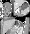
Figure 2. CT scan of the abdomen with IV contrast performed seven months earlier.
Coronal (A), axial (B) and sagittal (C) images show the gastric diverticulum (solid arrows). Note the air along the non-dependent aspect of the diverticulum on the axial (B) image (dashed arrow). The sagittal image (C) clearly shows a connection (open arrow) between the diverticulum (solid arrows) and the fundus of the stomach (dashed arrows).
Figure 3
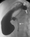
Figure 3. Upper GI exam performed on the same patient.
Fluoroscopic image of the stomach after the administration of barium with the patient in a right lateral decubitus position. There is contrast filling the gastric diverticulum (solid arrow) and the connection with the posterior aspect of the stomach (open arrow) can be seen.  The dashed arrow points to contrast emptying into the duodenum for orientation purposes.
Figure 4
Figure 4. Intraoperative view of the gastric diverticulum.

References

    1. Gastric diverticulum: a comprehensive review. Shah J, Patel K, Sunkara T, Papafragkakis C, Shahidullah A. Inflamm Intest Dis. 2018;3:161–166. - PMC - PubMed
    1. Gastric diverticula. Palmer ED. https://pubmed.ncbi.nlm.nih.gov/14840911/ Int Abstr Surg. 1951;92:417–428. - PubMed
    1. Surgical treatment of a gastric diverticulum in an adolescent. Elliott S, Sandler AD, Meehan JJ, Lawrence JP. J Pediatr Surg. 2006;41:1467–1469. - PubMed
    1. Gastric diverticulum: "a wayside house of ill fame" with a laparoscopic solution. DuBois B, Powell B, Voeller G. JSLS. 2012;16:473–477. - PMC - PubMed
    1. Laparoscopic resection of a large proximal gastric diverticulum. Fine A. Gastrointest Endosc. 1998;48:93–95. - PubMed

Publication types

LinkOut - more resources