

Osteochondral Injury, Management and Tissue Engineering Approaches
- PMID: 33251212
- PMCID: PMC7673409
- DOI: 10.3389/fcell.2020.580868
Osteochondral Injury, Management and Tissue Engineering Approaches
Abstract
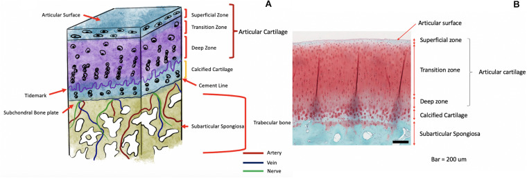
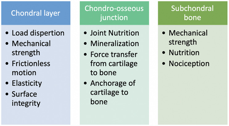
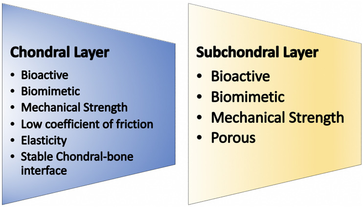
Osteochondral lesions (OL) are a common clinical problem for orthopedic surgeons worldwide and are associated with multiple clinical scenarios ranging from trauma to osteonecrosis. OL vary from chondral lesions in that they involve the subchondral bone and chondral surface, making their management more complex than an isolated chondral injury. Subchondral bone involvement allows for a natural healing response from the body as marrow elements are able to come into contact with the defect site. However, this repair is inadequate resulting in fibrous scar tissue. The second differentiating feature of OL is that damage to the subchondral bone has deleterious effects on the mechanical strength and nutritive capabilities to the chondral joint surface. The clinical solution must, therefore, address both the articular cartilage as well as the subchondral bone beneath it to restore and preserve joint health. Both cartilage and subchondral bone have distinctive functional requirements and therefore their physical and biological characteristics are very much dissimilar, yet they must work together as one unit for ideal joint functioning. In the past, the obvious solution was autologous graft transfer, where an osteochondral bone plug was harvested from a non-weight bearing portion of the joint and implanted into the defect site. Allografts have been utilized similarly to eliminate the donor site morbidity associated with autologous techniques and overall results have been good but both techniques have their drawbacks and limitations. Tissue engineering has thus been an attractive option to create multiphasic scaffolds and implants. Biphasic and triphasic implants have been under explored and have both a chondral and subchondral component with an interface between the two to deliver an implant which is biocompatible and emulates the osteochondral unit as a whole. It has been a challenge to develop such implants and many manufacturing techniques have been utilized to bring together two unalike materials and combine them with cellular therapies. We summarize the functions of the osteochondral unit and describe the currently available management techniques under study.
Keywords: Mesenchymal stem cell; Tissue Engineering and Regenerative Medicine; articular cartilage; multiphasic scaffold; osteochondral repair.
Copyright © 2020 Jacob, Shimomura and Nakamura.
Figures
Similar articles
-
Treatment of osteochondral defects in the rabbit's knee joint by implantation of allogeneic mesenchymal stem cells in fibrin clots.J Vis Exp. 2013 May 21;(75):e4423. doi: 10.3791/4423. J Vis Exp. 2013. PMID: 23728213 Free PMC article.
-
[Repairing defects of rabbit articular cartilage and subchondral bone with biphasic scaffold combined bone marrow stromal stem cells].Zhongguo Xiu Fu Chong Jian Wai Ke Za Zhi. 2010 Jan;24(1):87-93. Zhongguo Xiu Fu Chong Jian Wai Ke Za Zhi. 2010. PMID: 20135980 Chinese.
-
3D printing of fibre-reinforced cartilaginous templates for the regeneration of osteochondral defects.Acta Biomater. 2020 Sep 1;113:130-143. doi: 10.1016/j.actbio.2020.05.040. Epub 2020 Jun 4. Acta Biomater. 2020. PMID: 32505800
-
Osteochondral Tissue Engineering Dilemma: Scaffolding Trends in Regenerative Medicine.Stem Cell Rev Rep. 2023 Aug;19(6):1615-1634. doi: 10.1007/s12015-023-10545-x. Epub 2023 Apr 19. Stem Cell Rev Rep. 2023. PMID: 37074547 Review.
-
Autologous tissue transplantations for osteochondral repair.Dan Med J. 2016 Apr;63(4):B5236. Dan Med J. 2016. PMID: 27034191 Review.
Cited by
-
Human Chondrocytes from Human Adipose Tissue-Derived Mesenchymal Stem Cells Seeded on a Dermal-Derived Collagen Matrix Sheet: Our Preliminary Results for a Ready to Go Biotechnological Cartilage Graft in Clinical Practice.Stem Cells Int. 2021 Feb 23;2021:6664697. doi: 10.1155/2021/6664697. eCollection 2021. Stem Cells Int. 2021. PMID: 33679990 Free PMC article.
-
Tomographic Assessment of Bone Regeneration in Osteochondral Lesion Treated with Various Biomaterials in a Sheep Model Study.J Funct Biomater. 2025 Apr 1;16(4):120. doi: 10.3390/jfb16040120. J Funct Biomater. 2025. PMID: 40278228 Free PMC article.
-
Fabrication and Characterization of Bioactive Gelatin-Alginate-Bioactive Glass Composite Coatings on Porous Titanium Substrates.ACS Appl Mater Interfaces. 2022 Apr 6;14(13):15008-15020. doi: 10.1021/acsami.2c01241. Epub 2022 Mar 22. ACS Appl Mater Interfaces. 2022. PMID: 35316017 Free PMC article.
-
Treatment of Chondral Lesions in the Knee.Rev Bras Ortop (Sao Paulo). 2023 Aug 30;58(4):e551-e556. doi: 10.1055/s-0043-1772196. eCollection 2023 Aug. Rev Bras Ortop (Sao Paulo). 2023. PMID: 37663186 Free PMC article.
-
Additive-Free Gelatine-Based Devices for Chondral Tissue Regeneration: Shaping Process Comparison among Mould Casting and Three-Dimensional Printing.Polymers (Basel). 2022 Mar 4;14(5):1036. doi: 10.3390/polym14051036. Polymers (Basel). 2022. PMID: 35267859 Free PMC article.
References
-
- Ando W., Tateishi K., Hart D. A., Katakai D., Tanaka Y., Nakata K., et al. (2007). Cartilage repair using an in vitro generated scaffold-free tissue-engineered construct derived from porcine synovial mesenchymal stem cells. Biomaterials 28 5462–5470. 10.1016/j.biomaterials.2007.08.030 - DOI - PubMed
-
- Angele P., Schumann D., Angele M., Kinner B., Englert G., Hente R., et al. (2004). Cyclic, mechanical compression enhances chondrogenesis of mesenchymal progenitor cells in tissue engineering scaffolds. Biorheology 41 335–346. - PubMed
Publication types
LinkOut - more resources
Full Text Sources
Miscellaneous