. 2020:3:183.
doi: 10.1038/s42005-020-00449-4. Epub 2020 Oct 16.

Aspiration-assisted freeform bioprinting of prefabricated tissue spheroids in a yield-stress gel

Affiliations

Aspiration-assisted freeform bioprinting of prefabricated tissue spheroids in a yield-stress gel

Bugra Ayan et al. Commun Phys. 2020.

Abstract

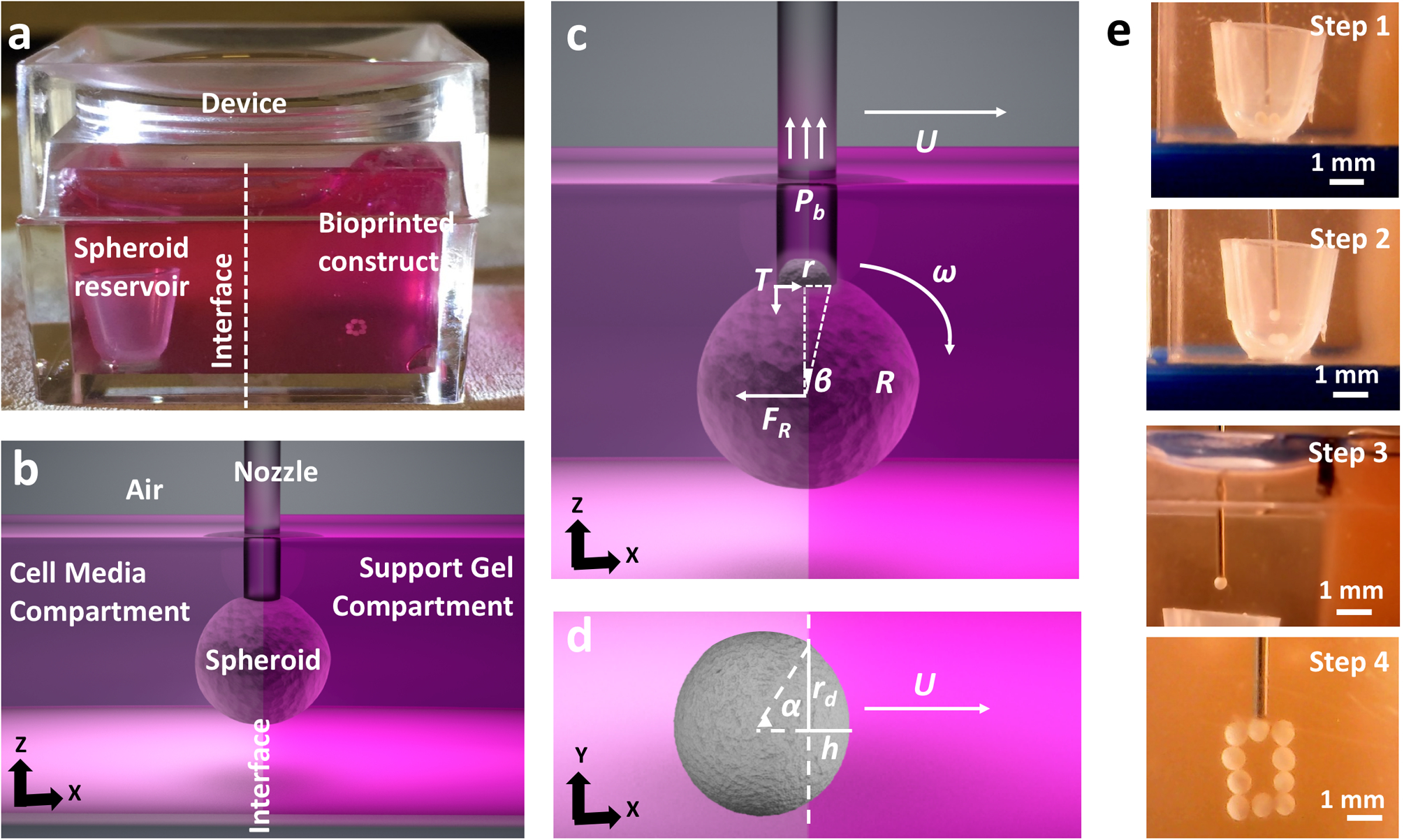
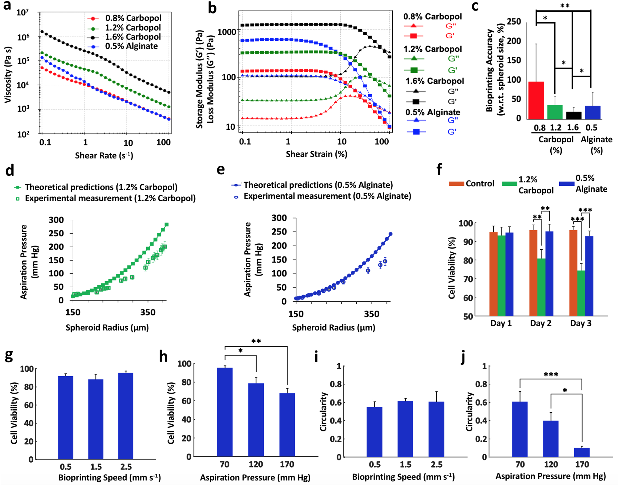
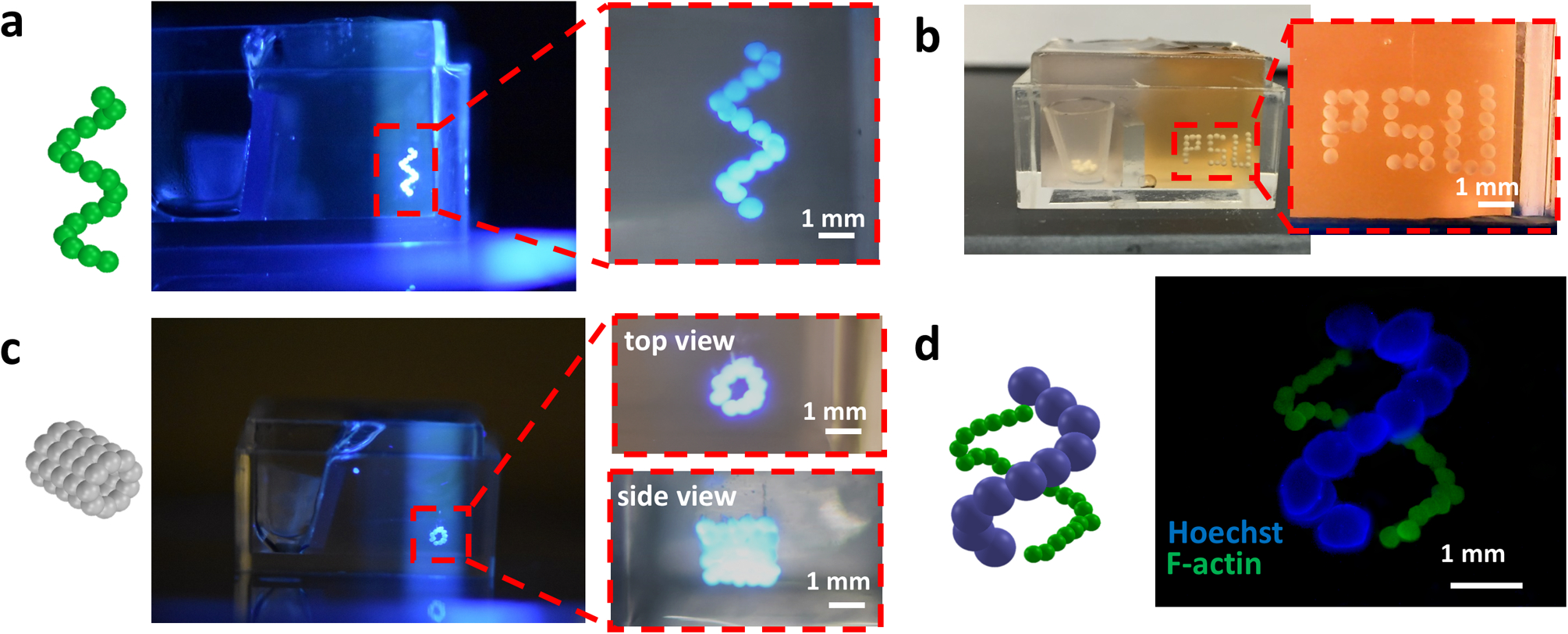
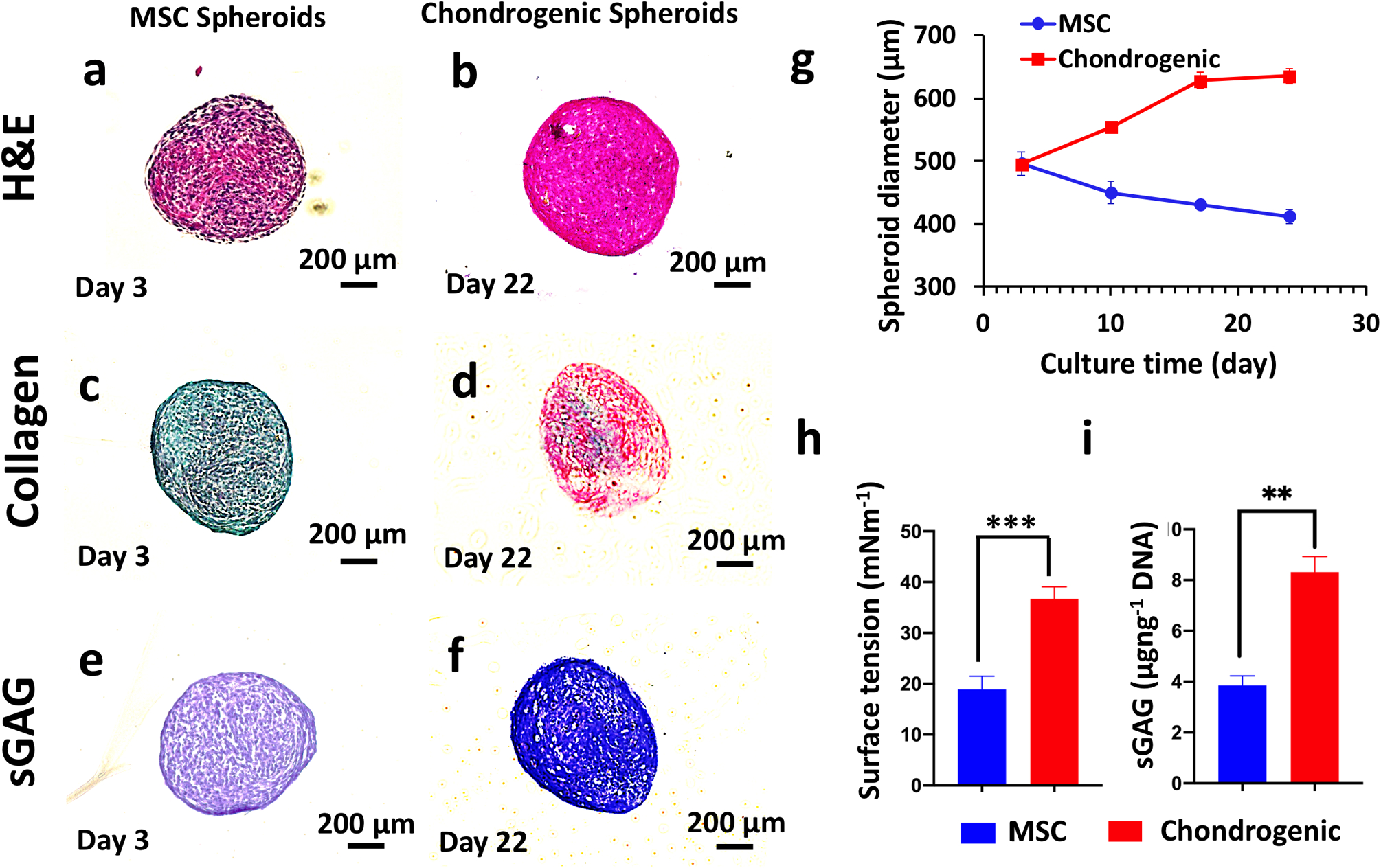
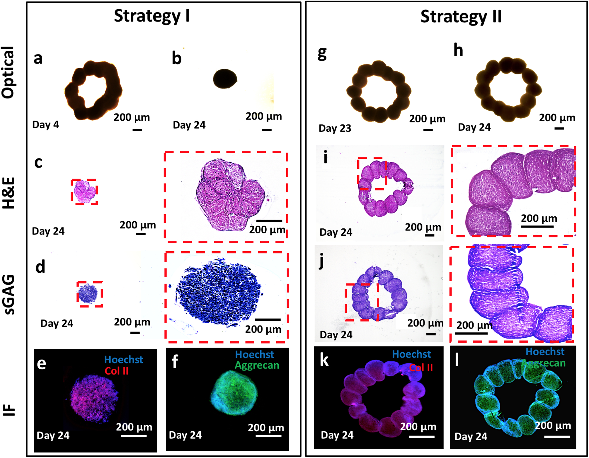
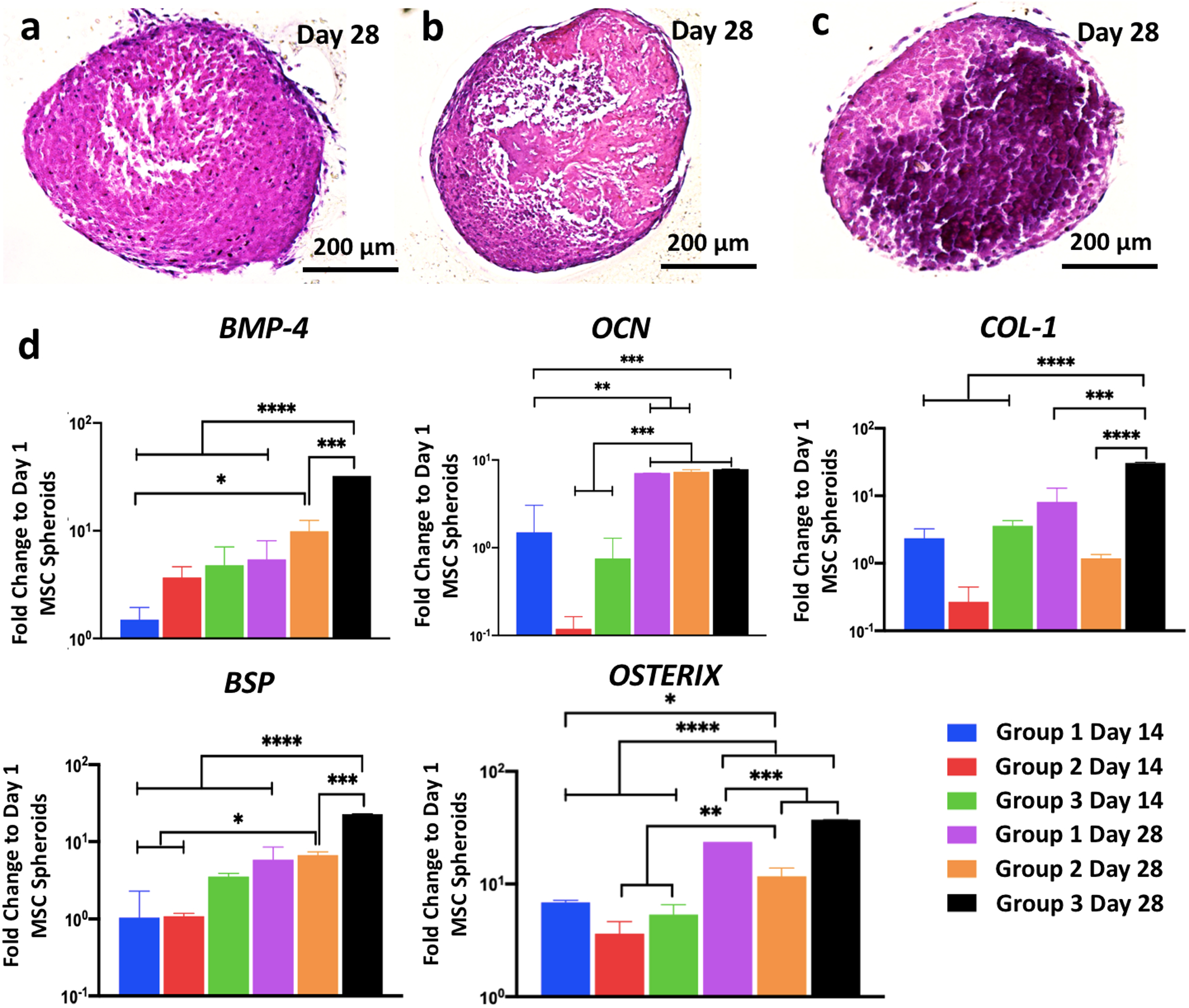
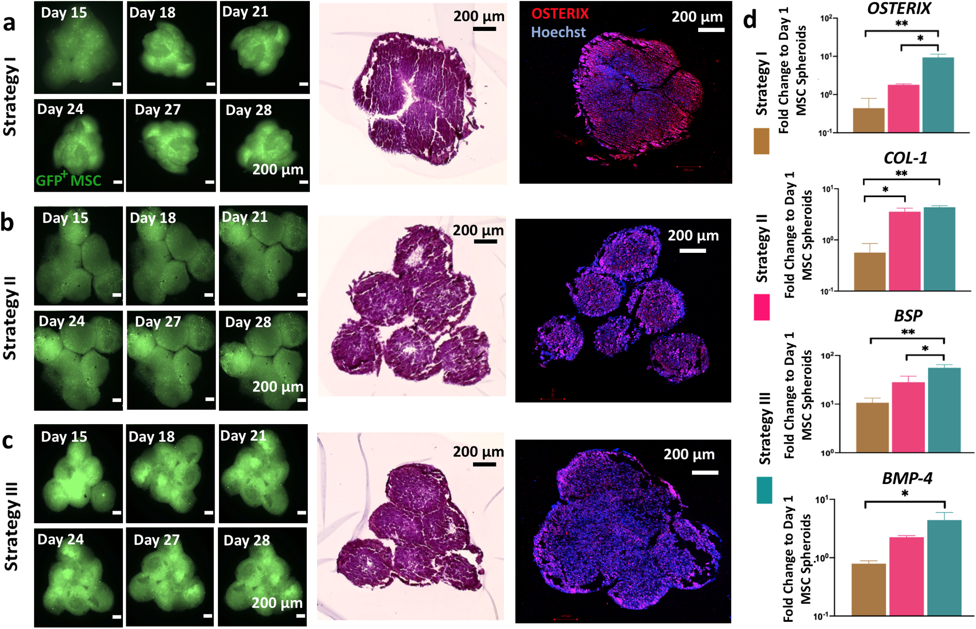
Bioprinting of cellular aggregates, such as tissue spheroids, to form three-dimensional (3D) complex-shaped arrangements, has posed a major challenge due to lack of robust, reproducible and practical bioprinting techniques. Here, we demonstrate 3D aspiration-assisted freeform bioprinting of tissue spheroids by precisely positioning them in self-healing yield-stress gels, enabling the self-assembly of spheroids for fabrication of tissues. The presented approach enables the traverse of spheroids directly from the cell media to the gel and freeform positioning of the spheroids on demand. We study the underlying physical mechanism of the approach to elucidate the interactions between the aspirated spheroids and the gel's yield-stress during the transfer of spheroids from cell media to the gel. We further demonstrate the application of the proposed approach in the realization of various freeform shapes and self-assembly of human mesenchymal stem cell spheroids for the construction of cartilage and bone tissues.


Conflict of interest statement

Competing interests The authors declare no competing interests.

Figures

Fig. 1
Fig. 1. Aspiration-assisted freeform bioprinting process.
a The bioprinting setup, where a box was filled with the yield-stress gel in one compartment and cell media in the other. b A schematic showing the process of spheroid traverse across the yield-stress gel and media compartment. c, d Schematics showing physical parameters involved in transferring of spheroids from the cell media to the yield-stress gel, FR is the magnitude of the resultant force acting on the spheroid due to its interaction with the environment, r is the nozzle’s radius, U is the bioprinting speed, and Pb, the critical aspiration pressure, is a function of R, r, U, gel properties (K, n, τ0). e Images showing a step-by-step illustration of the process, where (Step 1) spheroids were stored in the reservoir in the cell media, (Step 2) spheroids were picked from the reservoir, (Step 3) traversed the interface into yield-stress gel from the cell media compartment and (Step 4) bioprinted to form a predefined shape.
Fig. 2
Fig. 2. The interplay among gel, spheroid, and bioprinting process parameters and its role in spheroid viability and shape.
a, b Rheological properties of Carbopol at different concentrations and 0.5% alginate microparticles. The gels showed shear-thinning properties indicated by decreasing viscosity with shear rate. c The bioprinting accuracy of the yield-stress gels (with respect to spheroid size) (n = 5; *p < 0.05 and **p < 0.01). The positional precision for 0.8%, 1.2%, and 1.6% concentrations of Carbopol and 0.5% alginate microparticles were observed to be ~97%, 22%, 12%, and 34%, respectively. The colors correspond to the legend of panel (a). d, e Confirmation of the theoretical approach using the experimental validation for spheroids ranging from 150 to 450 μm in radius bioprinted in 1.2% Carbopol and 0.5% alginate microparticles. Note that human mesenchymal stem cell (MSC) spheroids were utilized in all experiments. The theoretical relation was plotted according to Eq. (6). f Cell viability of MSC spheroids in different yield-stress gels over 3 days (note that free standing MSC spheroids were used as a positive control, n = 3; **p < 0.01 and ***p < 0.001). gj Cell viability and circularity of MSC spheroids at different bioprinting speed and aspiration pressure in alginate microparticles (n = 3; *p < 0.05, **p < 0.01 and ***p < 0.001). Increasing the bioprinting speed from 0.5 to 2.5 mm s−1 did not reduce the cell viability when the aspiration pressure was maintained constant. However, increasing the aspiration pressure from 70 to 170 mm Hg decreased the cell viability. On the other hand, increase in the bioprinting speed did not significantly change the circularity of spheroids under a given aspiration pressure, whereas, increasing the aspiration pressure from 70 to 170 mm Hg increased the deformation of the spheroids and reduced their circularity. Error bars were plotted as mean ± standard deviation.
Fig. 3
Fig. 3. Aspiration-assisted freeform bioprinting of spheroids in different configurations.
Schematic illustration and optical photographs of 3D bioprinted a helix-shape (mesenchymal stem cell (MSC) spheroids), b initials of Penn State University (PSU, MSC spheroids), c five-layer tubular (MSC spheroids), and d double helix-shape constructs using MSC spheroids with 150 μm (F-actin) and 450 μm (Hoechst) in radius in 1.2% Carbopol yield-stress gel. The red dashed line denotes the region magnified.
Fig. 4
Fig. 4. Chondrogenic differentiation of mesenchymal stem cell spheroids.
Histological staining of mesenchymal stem cell (MSC) (at Day 3, prior to bioprinting for Strategy I) and chondrogenic spheroids (at Day 22, prior to bioprinting for Strategy II) a, b hematoxylin and eosin, c, d picrosirius red/fast green, and e, f toluidine blue staining. MSC spheroids were less dense and were negative (demonstrated by green color) for collagen and sGAG (demonstrated by purplish color), whereas the chondrogenic spheroids were denser and positive (demonstrated by green color) for collagen and sGAG (demonstrated by blue color). g Diameter change of MSC and chondrogenic spheroids over 24 days (n = 10). Note that chondrogenic spheroids were cultured in MSC growth media for the first 3 days of culture. The diameter of chondrogenic spheroids increased from 500 μm (on Day 3) to 600 μm (on Day 18) and retained their size for the remaining period of the culture until Day 24. The diameter of MSC spheroids gradually decreased from 500 μm (on Day 3) to 400 μm (on Day 24). h Surface tension, the surface tension values for both spheroids were within feasible ranges for bioprinting and i sGAG content measurements (normalized to DNA amount of MSC and chondrogenic spheroids at Day 24) (n = 3, **p < 0.01 and ***p < 0.001). A 2.2-fold increase in the sGAG content (μg ng−1 DNA) shown for chondrogenic spheroids as compared to MSC spheroids. Error bars were plotted as mean ± standard deviation.
Fig. 5
Fig. 5. Aspiration-assisted freeform bioprinting of circular cartilage tissues.
Strategy I: cartilages tissues were bioprinting using mesenchymal stem cell spheroids on Day 3 and removed from 0.5% alginate microparticles on Day 4. a A microscopic image showing a bioprinted construct after removal from alginate microparticles. b The final shape of the bioprinted cartilage at Day 24. Histological and immunostaining images of the bioprinted tissues at Day 24 including c hematoxylin and eosin (H&E), d toluidine blue, e Col-II, and f Aggrecan staining. Strategy II: cartilage tissues were bioprinted using chondrogenic spheroids at Day 22. g A microscopic image showing bioprinted construct after its removal from 0.5% alginate microparticles at Day 23. h The final shape of the bioprinted cartilage tissue at Day 24. Histological and immunostaining images of the bioprinted tissues at Day 24 including i H&E, j toluidine blue, k Col-II, and l Aggrecan. We showed that the bioprinted tissues in Strategy I exhibited chondrogenic properties; however, the bioprinted shape could not be retained because of the compaction of MSC spheroids. In Strategy II, we observed sufficient fusion between spheroids and the originally bioprinted circular arrangement was retained. The red dashed line denotes the region magnified.
Fig. 6
Fig. 6. Osteogenic differentiation of mesenchymal stem cell spheroids.
Hematoxylin and eosin staining images of spheroids of a Group 1, b Group 2, and c Group 3 at Day 28 demonstrating stronger bone matrix deposition in Group 3. d BMP-4, OCN, COL-1, BSP, and OSTERIX gene expressions of Group 1 Day 14, Group 2 Day 14, Group 3 Day 14, Group 1 Day 28, Group 2 Day 28, and Group 3 Day 28 (n = 3; *p < 0.05, **p < 0.01, ***p < 0.001, and ****p < 0.0001). All genes for all groups showed greater level of expression on Day 28 compared to Day 14. No significant difference was observed at Day 14 among groups; expression of BMP-4 (4.8- and 32.3-folds), COL-1 (3.6- and 30.5-folds), BSP (3.5- and 22.8-folds), and OSTERIX (5.3- and 37-folds) in Group 3 on Day 28 was significantly higher than those for Groups 1 and 2, respectively. Error bars have been plotted as mean ± standard deviation.
Fig. 7
Fig. 7. Aspiration-assisted freeform bioprinting of osteogenic tissues in a yield-stress gel.
Time-lapse images of green fluorescent protein (GFP)+ mesenchymal stem cell (MSC) spheroids and their immunostaining (Hoechst in blue and OSTERIX in red) and hematoxylin and eosin staining for bioprinted bone tissue using a Strategy I, b Strategy II, and c Strategy III. In Strategy I, the original shape could not be conserved due to compaction, whereas in Strategies II and III, the triangle shape was well preserved. d OSTERIX, COL-1, BSP, and BMP-4 gene expressions of 3D bioprinted bone tissues cultured using different strategies (n = 3; *p < 0.05, **p < 0.01, and ***p < 0.001). Constructs in Strategy III exhibited the highest expression level for all genes—23.5- and 5.2-fold increase for OSTERIX, 7.9- and 1.2-fold increase for COL-1, 5.2- and 2-fold increase for BSP, and 5.5- and 2-fold increase for BMP-4, respectively, compared to Strategies I and II. Error bars have been plotted as mean ± standard deviation.

Similar articles

Cited by

References

    1. Dababneh AB & Ozbolat IT Bioprinting technology: a current state-ofthe-art review. J. Manuf. Sci. Eng 136, 061016 (2014).
    1. Sun W et al. The bioprinting roadmap. Biofabrication 12, 022002 (2020). - PubMed
    1. Leberfinger AN et al. Bioprinting functional tissues. Acta Biomater 95, 32–49 (2019). - PMC - PubMed
    1. Ozbolat IT 3D Bioprinting: Fundamentals, Principles and Applications (Academic Press, London, 2016).
    1. Bhattacharjee T et al. Writing in the granular gel medium. Sci. Adv 1, e1500655 (2015). - PMC - PubMed