

Infectivity and pathogenicity of different hepatitis E virus genotypes/subtypes in rabbit model
- PMID: 33251979
- PMCID: PMC7781933
- DOI: 10.1080/22221751.2020.1858178
Infectivity and pathogenicity of different hepatitis E virus genotypes/subtypes in rabbit model
Abstract
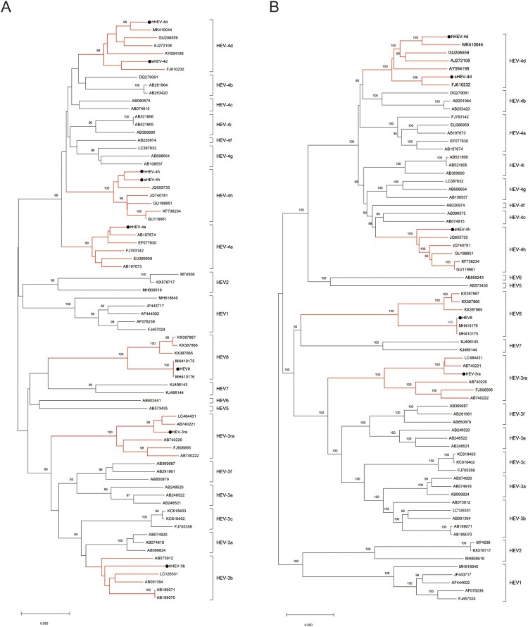
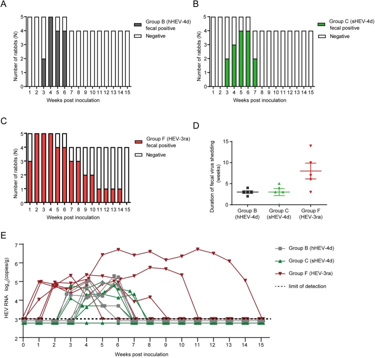
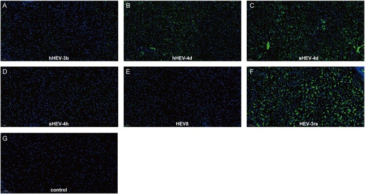
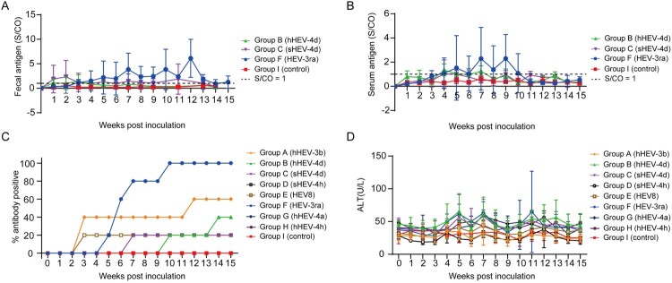
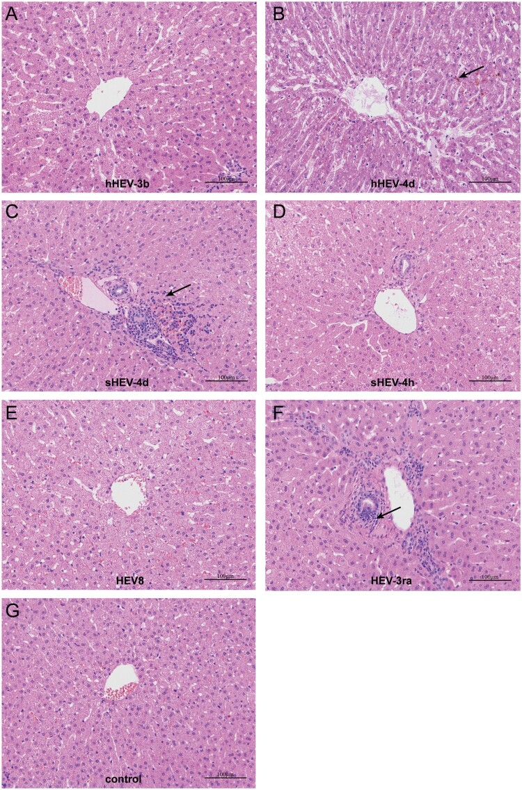
The pathogenicity of each hepatitis E virus (HEV) genotypes/subtypes may be different. This study aimed to investigate the infectivity and pathogenicity of different HEV genotypes/subtypes from different mammalian sources especially human in rabbits, and to assess whether rabbits are an appropriate animal model to study different HEV genotypes/subtypes. Thirty-seven rabbits were randomly divided into nine groups and inoculated with eight different HEV strains, including human-derived HEV3b (hHEV-3b), hHEV-4a, hHEV-4d and hHEV-4h, swine-derived HEV4d (sHEV-4d) and sHEV-4h, rabbit-derived HEV3 (HEV-3ra) and camel-derived HEV8. HEV RNA, antigen, anti-HEV and alanine aminotransferase (ALT) in serum or/and feces were monitored weekly. One rabbit from each group was euthanized at seven weeks post inoculation and the liver specimens were taken for histopathological analysis and immunofluorescence staining of HEV ORF2 proteins. hHEV-4d, sHEV-4d and HEV-3ra infections were successfully established in rabbits and typical acute hepatitis symptoms were observed, including viraemia/antigenemia, fecal virus/antigen shedding, elevated ALT level and liver histopathological changes. One rabbit infected with HEV-3ra showed chronic infection. hHEV-4d and sHEV-4d are less infectious and pathogenic than HEV-3ra in rabbits. hHEV-3b and HEV8 only caused inapparent infection in rabbits as 60% (3/5) and 20% (1/5) of the rabbits seroconverted to anti-HEV, respectively. No obvious signs of HEV infection in rabbits inoculated with hHEV-4a, hHEV-4h and sHEV-4h. The infectivity and pathogenicity of different HEV genotypes/subtypes in rabbits is different, which may be related to the species specificity of HEV. Rabbit can be used as an animal model for the study of HEV-3ra and more importantly human HEV-4d.
Keywords: Hepatitis E virus; infectivity; pathogenicity; rabbit model; susceptibility.
Conflict of interest statement
No potential conflict of interest was reported by the author(s).
Figures
Similar articles
-
Experimental infection of rabbits with rabbit and genotypes 1 and 4 hepatitis E viruses.PLoS One. 2010 Feb 11;5(2):e9160. doi: 10.1371/journal.pone.0009160. PLoS One. 2010. PMID: 20161794 Free PMC article.
-
Different susceptibility and pathogenesis of rabbit genotype 3 hepatitis E virus (HEV-3) and human HEV-3 (JRC-HE3) in SPF rabbits.Vet Microbiol. 2017 Aug;207:1-6. doi: 10.1016/j.vetmic.2017.05.019. Epub 2017 May 29. Vet Microbiol. 2017. PMID: 28757007
-
Experimental infection of rabbit with swine-derived hepatitis E virus genotype 4.Vet Microbiol. 2019 Feb;229:168-175. doi: 10.1016/j.vetmic.2019.01.001. Epub 2019 Jan 4. Vet Microbiol. 2019. PMID: 30642594
-
Hepatitis E Virus Genotypes and Evolution: Emergence of Camel Hepatitis E Variants.Int J Mol Sci. 2017 Apr 20;18(4):869. doi: 10.3390/ijms18040869. Int J Mol Sci. 2017. PMID: 28425927 Free PMC article. Review.
-
An overview: Rabbit hepatitis E virus (HEV) and rabbit providing an animal model for HEV study.Rev Med Virol. 2018 Jan;28(1). doi: 10.1002/rmv.1961. Epub 2017 Nov 17. Rev Med Virol. 2018. PMID: 29148605 Review.
Cited by
-
Molecular epidemiology and genotype-specific disease severity of hepatitis E virus infections in Germany, 2010-2019.Emerg Microbes Infect. 2022 Dec;11(1):1754-1763. doi: 10.1080/22221751.2022.2091479. Emerg Microbes Infect. 2022. PMID: 35713010 Free PMC article.
-
Ribavirin Treatment Failure-Associated Mutation, Y1320H, in the RNA-Dependent RNA Polymerase of Genotype 3 Hepatitis E Virus (HEV) Enhances Virus Replication in a Rabbit HEV Infection Model.mBio. 2023 Apr 25;14(2):e0337222. doi: 10.1128/mbio.03372-22. Epub 2023 Feb 21. mBio. 2023. PMID: 36809085 Free PMC article.
-
Prevalence of Hepatitis E Virus Infection among Laboratory Rabbits in China.Pathogens. 2021 Jun 21;10(6):780. doi: 10.3390/pathogens10060780. Pathogens. 2021. PMID: 34205738 Free PMC article.
-
A small animal model of chronic hepatitis E infection using immunocompromised rats.JHEP Rep. 2022 Aug 10;4(10):100546. doi: 10.1016/j.jhepr.2022.100546. eCollection 2022 Oct. JHEP Rep. 2022. PMID: 36052220 Free PMC article.
-
Immunisation of pigs with recombinant HEV vaccines does not protect from infection with HEV genotype 3.One Health. 2024 Jan 6;18:100674. doi: 10.1016/j.onehlt.2023.100674. eCollection 2024 Jun. One Health. 2024. PMID: 39010962 Free PMC article.
References
-
- Kamar N, Izopet J, Pavio N, et al. . Hepatitis E virus infection. Nat Rev Dis Primers. 2017;3:17086. - PubMed
-
- Patra S, Kumar A, Trivedi SS, et al. . Maternal and fetal outcomes in pregnant women with acute hepatitis E virus infection. Ann Intern Med. 2007;147(1):28–33. - PubMed
-
- Perez-Gracia MT, Suay-Garcia B, Mateos-Lindemann ML.. Hepatitis E and pregnancy: current state. Rev Med Virol. 2017;27(3):e1929. - PubMed
-
- Kamar N, Mansuy JM, Cointault O, et al. . Hepatitis E virus-related cirrhosis in kidney- and kidney-pancreas-transplant recipients. Am J Transpl. 2008;8(8):1744–1748. - PubMed
MeSH terms
Substances
LinkOut - more resources
Full Text Sources