. 2020 Dec;131(1):e114.
doi: 10.1002/cpim.114.

Protocols for Experimental Sjögren's Syndrome

Affiliations

Protocols for Experimental Sjögren's Syndrome

Harini Bagavant et al. Curr Protoc Immunol. 2020 Dec.

Abstract

Sjögren's syndrome (SS) is a systemic autoimmune disease affecting multiple organ systems. Salivary and lacrimal gland involvement cause dry mouth and dry eye and are the most common clinical presentations of the disease. Patients with SS also have autoantibodies targeting multiple nuclear and cytoplasmic antigens. Innate immune activation plays a critical role in SS pathogenesis. This article describes the activation of specific innate immune pathways in mice to study SS salivary gland manifestations. Methodologies for evaluating salivary gland inflammation and salivary function are described. This article also describes protocols for in-house assays to measure autoantibody titers in serum. © 2020 Wiley Periodicals LLC Basic Protocol 1: Acceleration of Sjögren's syndrome by activating the toll-like receptor 3 pathway Basic Protocol 2: Induction of Sjögren's syndrome by activating the stimulator of interferon genes pathway Alternate Protocol: Acceleration of Sjögren's syndrome by the administration of Freund's incomplete adjuvant Support Protocol 1: Evaluating salivary gland function Support Protocol 2: Evaluating salivary gland inflammation Support Protocol 3: Measuring autoantibody titers by indirect immunofluorescence.

Keywords: STING; Sjögren's syndrome; TLR3; innate immunity; salivary glands.


Conflict of interest statement

CONFLICT OF INTEREST: The authors declare no conflicts of interest.

Figures

Figure 1:
Figure 1:
Schematic representation of the timeline for Basic protocol 1 (A) and Basic Protocol 2 (B).
Figure 2:
Figure 2:
Dissection of salivary glands from mice. (A) Give a midline incision from the sternum to the lower lip’s base and raise the skin flaps on either side to expose the salivary glands. (B) Identify the submandibular (left SMG- black outline), sublingual (left SLG - blue outline), and parotid (left PG – green outline) glands. Note that the cervical lymph nodes (CxLN – yellow dots) are embedded in this area and may be visible on dissection.
Figure 3:
Figure 3:
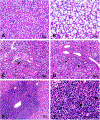
Evaluating salivary gland inflammation. Photomicrographs of formalin-fixed, paraffin-embedded, hematoxylin and eosin-stained mouse salivary glands showing normal ductal and acinar organization in SMG (A) and SLG (B). SMG showing a small, single lymphocytic focus (arrow) in the peri-vascular, peri-ductal region (C), and a larger lymphocytic focus invading the surrounding acinar tissues (arrows) (D). Extensive lymphocytic infiltration of the SMG (E), with lymphocytes invading the glandular parenchyma (F) showing remnants of acinar and ductal tissue (arrows). Objectives used for capturing the Images A-D (20x), E (10x) and F (40x). Scale bar = 50 microns.
Figure 4:
Figure 4:
Quantitative evaluation of salivary gland inflammation. (A) The digitally scanned image of a cross-section through all the salivary glands harvested together showing SMG, SLG, and PG. (B) Areas covered by lymphocytic foci (n=17, green outline ) and the total area of the section (blue outline). Severity of inflammation for this sample may be reported as (i) Area of inflammation (%) = area with lymphocytic infiltration (0.956 mm2)/ total area of the section (23.379 mm2) *100 = 4.09%; or (ii) Focus scores = [no. of foci (17)/ total area of the section (23.379)] *4 = 2.91.
Figure 5:
Figure 5:
Autoantibody detection by indirect immunofluorescence. HeLa cells grown on coverslips were probed with sera from DMXAA injected mice (A-H) or control mouse sera (I-J). Bound antibodies were detected with goat anti-mouse IgG-FITC (Green), and nuclei were stained with DAPI (blue). Panel K-L show staining with only secondary antibody. Sera from DMXAA treated mice show weak (A, B) or strong (C, D) homogenous nuclear staining. Panels (E, F) show cytoplasmic staining, and (G, H) show prominent nuclear membrane staining. All the sera were tested at 1:50 dilution, and images were captured at 20x on a Zeiss LSM-710 confocal microscope. Scale bar = 20 microns.

References

    1. Allushi B, Bagavant H, Papinska J, & Deshmukh US (2019). Hyperglycemia and Salivary Gland Dysfunction in the Non-obese Diabetic Mouse: Caveats for Preclinical Studies in Sjögren’s Syndrome. Scientific reports, 9(1), 17969. - PMC - PubMed
    1. Amador-Patarroyo MJ, Arbelaez JG, Mantilla RD, Rodriguez-Rodriguez A, Cárdenas-Roldán J, Pineda-Tamayo R, Guarin MR, Kleine LL, Rojas-Villarraga A, & Anaya JM (2012). Sjögren’s syndrome at the crossroad of polyautoimmunity. Journal of autoimmunity, 39(3), 199–205. - PubMed
    1. Bagavant H, Trzeciak M, Papinska J, Biswas I, Dunkleberger ML, Sosnowska A, & Deshmukh US (2018). A Method for the Measurement of Salivary Gland Function in Mice. Journal of visualized experiments : JoVE, (131), 57203. - PMC - PubMed
    1. Cha S, Nagashima H, Brown VB, Peck AB, & Humphreys-Beher MG (2002). Two NOD Idd-associated intervals contribute synergistically to the development of autoimmune exocrinopathy (Sjögren’s syndrome) on a healthy murine background. Arthritis and rheumatism, 46(5), 1390–1398. - PubMed
    1. Chan EK, Damoiseaux J, Carballo OG, Conrad K, de Melo Cruvinel W, Francescantonio PL, Fritzler MJ, Garcia-De La Torre I, Herold M, Mimori T, Satoh M, von Mühlen CA, & Andrade LE (2015). Report of the First International Consensus on Standardized Nomenclature of Antinuclear Antibody HEp-2 Cell Patterns 2014–2015. Frontiers in immunology, 6, 412. 10.3389/fimmu.2015.00412 - DOI - PMC - PubMed

Publication types