. 2020 Nov 30;16(11):e1009025.
doi: 10.1371/journal.ppat.1009025. eCollection 2020 Nov.

TLR-9 agonist and CD40-targeting vaccination induces HIV-1 envelope-specific B cells with a diversified immunoglobulin repertoire in humanized mice

Affiliations

TLR-9 agonist and CD40-targeting vaccination induces HIV-1 envelope-specific B cells with a diversified immunoglobulin repertoire in humanized mice

Véronique Godot et al. PLoS Pathog. .

Abstract

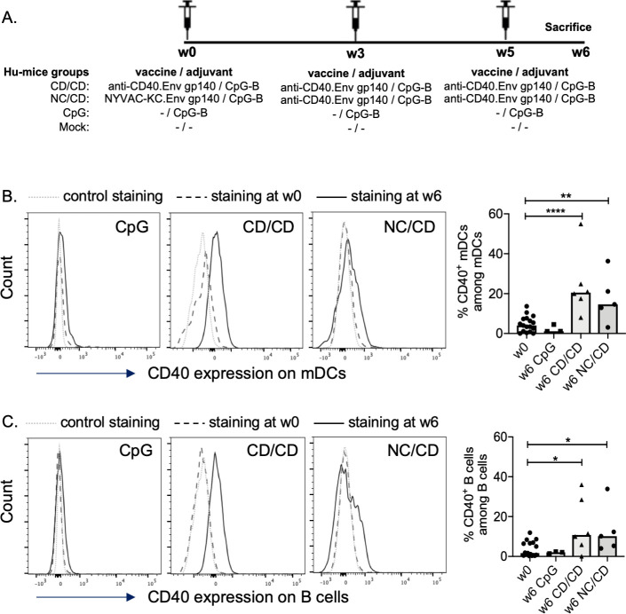
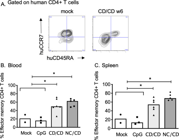
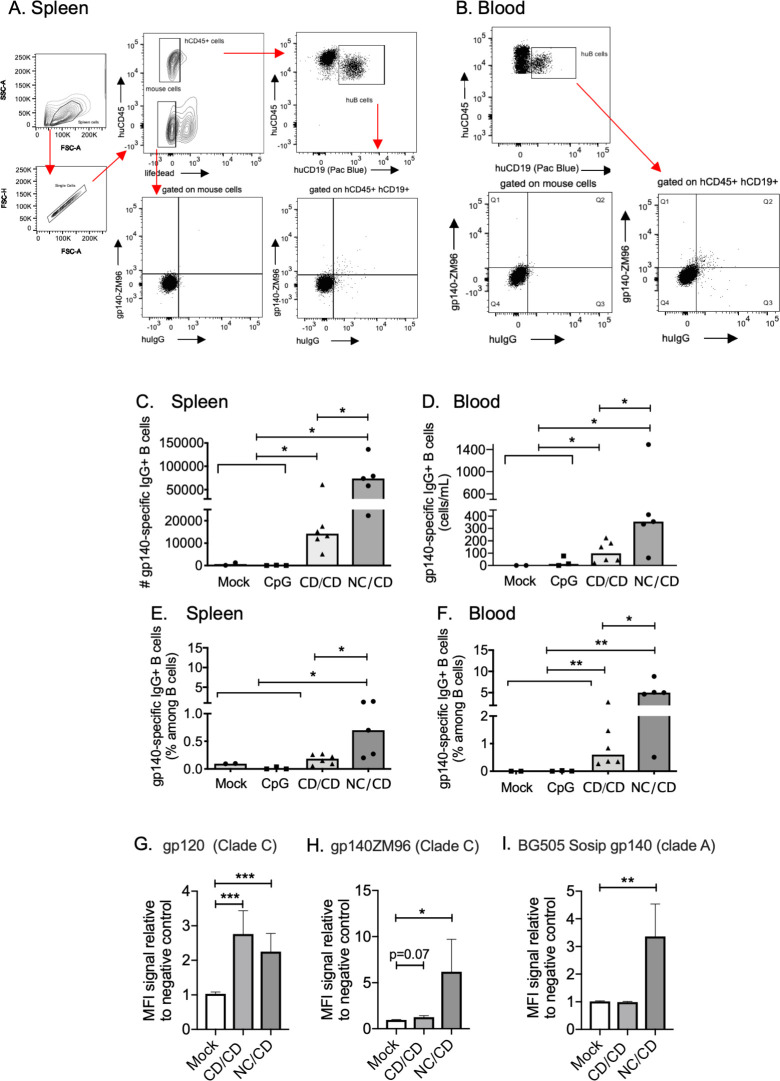
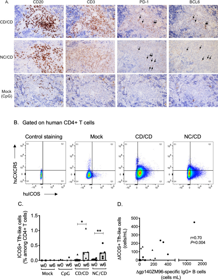
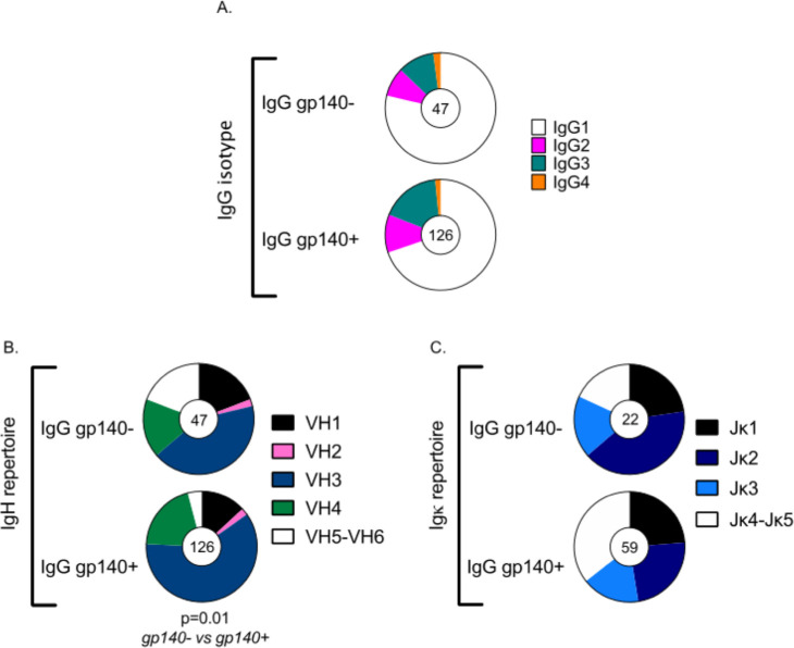
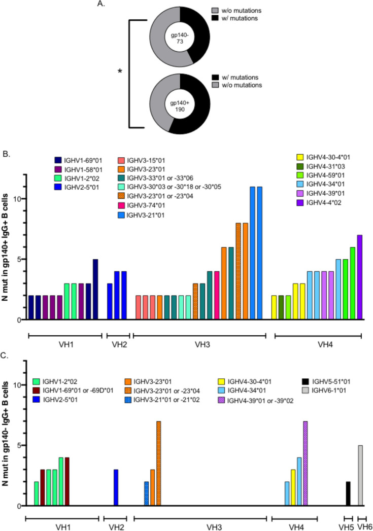
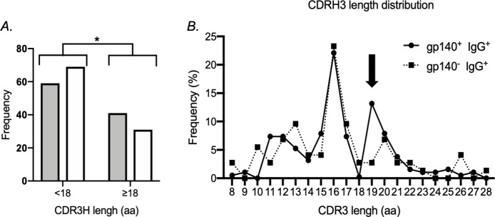
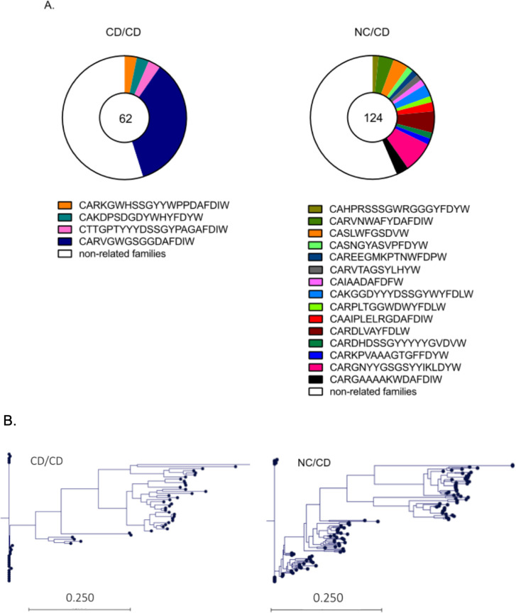
The development of HIV-1 vaccines is challenged by the lack of relevant models to accurately induce human B- and T-cell responses in lymphoid organs. In humanized mice reconstituted with human hematopoietic stem cells (hu-mice), human B cell-development and function are impaired and cells fail to efficiently transition from IgM B cells to IgG B cells. Here, we found that CD40-targeted vaccination combined with CpG-B adjuvant overcomes the usual defect of human B-cell switch and maturation in hu-mice. We further dissected hu-B cell responses directed against the HIV-1 Env protein elicited by targeting Env gp140 clade C to the CD40 receptor of antigen-presenting cells. The anti-CD40.Env gp140 vaccine was injected with CpG-B in a homologous prime/boost regimen or as a boost of a NYVAC-KC pox vector encoding Env gp140 clade C. Both regimens elicited Env-specific IgG-switched memory hu-B cells at a greater magnitude in hu-mice primed with NYVAC-KC. Single-cell RNA-seq analysis showed gp140-specific hu-B cells to express polyclonal IgG1 and IgG3 isotypes and a broad Ig VH/VL repertoire, with predominant VH3 family gene usage. These cells exhibited a higher rate of somatic hypermutation than the non-specific IgG+ hu-B-cell counterpart. Both vaccine regimens induced splenic GC-like structures containing hu-B and hu-Tfh-like cells expressing PD-1 and BCL-6. We confirmed in this model that circulating ICOS+ memory hu-Tfh cells correlated with the magnitude of gp140-specific B-cell responses. Finally, the NYVAC-KC heterologous prime led to a more diverse clonal expansion of specific hu-B cells. Thus, this study shows that CD40-targeted vaccination induces human IgG production in hu-mice and provides insights for the development of a CD40-targeting vaccine to prevent HIV-1 infection in humans.


Conflict of interest statement

I have read the journal's policy and the authors of this manuscript have the following competing interests: GZ, SZ, and YL are named inventors on CD40-targeting vaccine patents and patent filings held jointly by INSERM and the Baylor Research Institute.

Figures

Fig 1
Fig 1. Enhanced expression of CD40 on myeloid (m) human DCs and B cells in vaccinated hu-mice.
(A) Schematic representation of the experimental design. (B-C) Representative histograms and summary data show the frequency of human blood CD40+ mDCs (B) and human CD40+ B lymphocytes (C) in the different vaccination settings. See the gating strategy of human mDCs and B cells in the S1 Fig. Individual values are presented, along with the median. The values of all hu-mice at week 0 was used as the base values. Mann-Whitney U-tests were used for comparisons. *p < 0.05, **p < 0.01, ****p<0.0001.
Fig 2
Fig 2. Vaccination elicits expansion of human memory T cells.
(A) Flow cytometry of splenocytes from hu-mice injected three times with PBS (mock) or the anti-CD40.Env gp140 vaccine (CD/CD) one week after the last injection (w6). The human CD4+ T cells (see their gating strategy in S1 Fig) were represented in a huCCR7 versus huCD45RA dot blot to identify the human effector memory CD4+ T cells (CCR7- CD45RA-). (B-C) Frequency of human effector memory CD4+ T cells in the blood (B) and spleens (C) of hu-mice. Individual values are presented, along with the median. Mann-Whitney U-tests were used for comparisons between immunized and non-immunized hu-mice. *p < 0.05.
Fig 3
Fig 3. The CD/CD and NC/CD vaccination regimens elicit gp140ZM96 (Env)-specific human B cells.
(A-B) Gating strategy to identify the gp140ZM96-specific IgG+ human B cells (called gp140+). (A) Flow cytometry of splenocytes from concatenated NC/CD hu-mice cells one week after the last injection (w6). After gating for single cells, viable cells within the huCD45+ gate were represented in a huCD45 versus huCD19 dot blot. Then hu-B cells were represented in a gp140ZM96 versus huIgG dot blot. The control staining for gp140ZM96 and huIgG are showed by gating on mouse cells. (B) The same gating strategy was used to identify the blood gp140+-specific IgG+ human B cells from concatenated NC/CD hu-mice cells one week after the last injection (w6). (C-D) Total number of gp140+ IgG+ human B cells in the spleens (C) and blood (D) of hu-mice. (E-F) Frequencies of gp140+ IgG+ human B cells in the spleens (E) and blood (F) of hu-mice. (C to F) Individual values are presented, along with the median. (G to I) Plasma HIV-1-specific human antibodies from hu-mouse samples evaluated for HIV-1 gp120 (G), gp140ZM96 (H) and BG505 SOSIP gp140 (I) binding by a custom multiplex assay (mean +/- sem). Three independent determinations were performed in triplicate. Mann-Whitney U-tests were used for comparisons. *p < 0.05, **p<0.01, ***p<0.001.
Fig 4
Fig 4. The CD/CD and NC/CD vaccination regimens elicit human memory Tfh cells that correlate with the expansion of gp140-specific human B cells.
(A) Representative immunohistochemistry images of spleen sections from vaccinated hu-mice (NC/CD and CD/CD groups) and control animals that received CpG-B only (mock group). (B) Gating strategy for the evaluation of circulating human memory Tfh cells. Dot blots show concatenated data from mock hu-mice (n = 5), CD/CD (n = 6) or NC/CD (n = 5) vaccinated hu-mice one week after the last injection (w6). The human CD4+ T cells (see their gating strategy in the S1 Fig) were represented in a huCXCR5 versus huICOS dot blot to identify the subset of human memory blood ICOS+ Tfh cells. Control of hCXCR5 and hICOS staining was obtained by gating on mouse cells. (C) Frequency of human ICOS+ CXCR5+ Tfh cells in the blood of hu-mice. Individual values are presented, along with the median. Mann-Whitney U-tests were used for comparisons between w0 and w6 values obtained in immunized hu-mice. *p < 0.05, **p < 0.01. (D) Correlation between the increase in blood gp140-specific IgG+ hu-B cells (x axis) and the increase in blood ICOS+CXCR5+ hu-Tfh cells; hu-mice from the NC/CD group are represented as circles, hu-mice from the CD/CD group as triangles, and control groups as squares. Spearman’s rank test was used to assess correlations.
Fig 5
Fig 5. Immunoglobulin gene (IgH and IgL) repertoire of human gp140+ and gp140- IgG+ B cells in vaccinated hu-mice.
(A) Distribution of IgG subtypes within gp140+ and gp140- IgG+ human B cells in all immunized hu-mice. (B) VH usage of the IgH and (C) Jk usage of the IgL in gp140+ and gp140- IgG+ hu-B cells for all immunized hu-mice. The number of analyzed cells is indicated in the center of the pie charts. Distributions were compared using Chi-Square tests. The analysis of gene segment usage was carried out using NCBI IgBLAST software (http://www.ncbi.nlm.nih.gov/igblast/).
Fig 6
Fig 6. Mutated immunoglobulin VH gene status of human gp140+ and gp140- IgG+ B cells in vaccinated hu-mice.
(A) Frequencies of human gp140+-specific and non-specific (gp140-) IgG+ B cells, with or without mutations. The number of analyzed human cells is indicated in the center of the pie charts. Distributions were compared using Chi-Square tests. *p < 0.05. (B-C) Analysis of heavy chain gene segment usage and the number of somatic mutations in VH segments, carried out using NCBI IgBLAST software (http://www.ncbi.nlm.nih.gov/igblast/), in gp140+ (B) and gp140- (C) IgG+ hu-B cells from all immunized hu-mice. Only IgG+ B cells with more than one mutation are represented.
Fig 7
Fig 7. Characteristics of Complementarity Determining Region 3 of IgH (CDRH3) of human gp140+ and gp140- IgG+ B cells in vaccinated hu-mice.
(A-B) Comparison of the CDRH3 amino-acid length between gp140+ specific and non-specific (gp140-) IgG+ antibodies of all immunized hu-mice. (A) Distribution of gp140+ (grey bar) and gp140- (white bar) IgG+ hu-B cells into two categories depending on whether the length of their CDRH3 was < 18 amino acids or ≥18 amino acids. Distributions were compared using Chi-Square tests. *p < 0.05. (B) CDRH3 length distribution in gp140+ (solid line) and gp140- (dotted line) IgG+ hu-B cells of all immunized hu-mice. The arrow highlights the main difference between the two populations of IgG+ human B cells.
Fig 8
Fig 8. Antibody clonal families and their phylogeny within gp140+-specific IgG+ hu-B cells.
(A) Pie charts of clonal family representation were constructed from the IgH sequences and CDR3 segment of sorted gp140+-specific IgG+ hu-B cells. The white slice represents sequences that appeared only once. The colored slices represent clonally related sequences, for which each color accounts for a different IgH clone. The numbers in the pie charts represent the number of mAbs evaluated. Human B cells were considered to belong to the same clone on the basis of identical V, D, and J gene segment usage and CDR3 length for both heavy- and light-chain Ig genes. (B) Phylogenic trees were generated after sequence alignment using CLC Main Workbench 7 software (v7.0) with default parameters. The relationship between sequences was determined via the neighbor-joining method. Each tip represents a BCR heavy chain sequence. The scale bar represents the genetic distance (expected changes per nucleotide site).

References

    1. Flynn NM, Forthal DN, Harro CD, Judson FN, Mayer KH, Para MF, et al. Placebo-controlled phase 3 trial of a recombinant glycoprotein 120 vaccine to prevent HIV-1 infection. J Infect Dis. 2005;191(5):654–65. 10.1086/428404 - DOI - PubMed
    1. Paterson JC, Ballabio E, Mattsson G, Turner SH, Mason DY, Marafioti T. Labeling of multiple cell markers and mRNA using automated apparatus. Appl Immunohistochem Mol Morphol. 2008;16(4):371–81. 10.1097/PAI.0b013e318164fc63 - DOI - PubMed
    1. Buchbinder SP, Mehrotra DV, Duerr A, Fitzgerald DW, Mogg R, Li D, et al. Efficacy assessment of a cell-mediated immunity HIV-1 vaccine (the Step Study): a double-blind, randomised, placebo-controlled, test-of-concept trial. Lancet. 2008;372(9653):1881–93. 10.1016/S0140-6736(08)61591-3 - DOI - PMC - PubMed
    1. Gray GE, Allen M, Moodie Z, Churchyard G, Bekker LG, Nchabeleng M, et al. Safety and efficacy of the HVTN 503/Phambili study of a clade-B-based HIV-1 vaccine in South Africa: a double-blind, randomised, placebo-controlled test-of-concept phase 2b study. Lancet Infect Dis. 2011;11(7):507–15. 10.1016/S1473-3099(11)70098-6 - DOI - PMC - PubMed
    1. Hammer SM, Sobieszczyk ME, Janes H, Karuna ST, Mulligan MJ, Grove D, et al. Efficacy trial of a DNA/rAd5 HIV-1 preventive vaccine. N Engl J Med. 2013;369(22):2083–92. 10.1056/NEJMoa1310566 - DOI - PMC - PubMed

Publication types