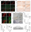
BMP5/7 protect dopaminergic neurons in an α-synuclein mouse model of Parkinson's disease
- PMID: 33253359
- PMCID: PMC7940172
- DOI: 10.1093/brain/awaa368
BMP5/7 protect dopaminergic neurons in an α-synuclein mouse model of Parkinson's disease
Figures


Comment in
-
Reply: Growth differentiation factor 5 exerts neuroprotection in an α-synuclein rat model of Parkinson's disease and BMP5/7 protect dopaminergic neurons in an α-synuclein mouse model of Parkinson's disease.Brain. 2021 Mar 3;144(2):e16. doi: 10.1093/brain/awaa369. Brain. 2021. PMID: 33257974 No abstract available.
Comment on
-
Randomized trial of intermittent intraputamenal glial cell line-derived neurotrophic factor in Parkinson's disease.Brain. 2019 Mar 1;142(3):512-525. doi: 10.1093/brain/awz023. Brain. 2019. PMID: 30808022 Free PMC article. Clinical Trial.
Similar articles
-
Endogenous alpha-synuclein influences the number of dopaminergic neurons in mouse substantia nigra.Exp Neurol. 2013 Oct;248:541-5. doi: 10.1016/j.expneurol.2013.07.015. Epub 2013 Aug 8. Exp Neurol. 2013. PMID: 23933574 Free PMC article.
-
Analyzing the Parkinson's Disease Mouse Model Induced by Adeno-associated Viral Vectors Encoding Human α-Synuclein.J Vis Exp. 2022 Jul 29;(185). doi: 10.3791/63313. J Vis Exp. 2022. PMID: 35969046
-
ZSCAN21 mediates the pathogenic transcriptional induction of α-synuclein in cellular and animal models of Parkinson's disease.Cell Death Dis. 2025 May 16;16(1):394. doi: 10.1038/s41419-025-07722-w. Cell Death Dis. 2025. PMID: 40379611 Free PMC article.
-
Cellular models for Parkinson's disease.J Neurochem. 2016 Oct;139 Suppl 1:121-130. doi: 10.1111/jnc.13618. Epub 2016 Apr 18. J Neurochem. 2016. PMID: 27091001 Review.
-
Parkinson's Disease: Exploring Different Animal Model Systems.Int J Mol Sci. 2023 May 22;24(10):9088. doi: 10.3390/ijms24109088. Int J Mol Sci. 2023. PMID: 37240432 Free PMC article. Review.
Cited by
-
Selective deletion of zinc transporter 3 in amacrine cells promotes retinal ganglion cell survival and optic nerve regeneration after injury.Neural Regen Res. 2023 Dec;18(12):2773-2780. doi: 10.4103/1673-5374.373660. Neural Regen Res. 2023. PMID: 37449644 Free PMC article.
-
Alpha-synuclein-induced upregulation of SKI family transcriptional corepressor 1: A new player in aging and Parkinson's disease?Neural Regen Res. 2026 Jan 1;21(1):320-321. doi: 10.4103/NRR.NRR-D-24-01156. Epub 2024 Dec 16. Neural Regen Res. 2026. PMID: 39688564 Free PMC article. No abstract available.
-
Key Connectomes and Synaptic-Compartment-Specific Risk Genes Drive Pathological α-Synuclein Spreading.Adv Sci (Weinh). 2025 Jul;12(25):e2413052. doi: 10.1002/advs.202413052. Epub 2025 May 28. Adv Sci (Weinh). 2025. PMID: 40433888 Free PMC article.
-
Functional Differentiation of BMP7 Genes in Zebrafish: bmp7a for Dorsal-Ventral Pattern and bmp7b for Melanin Synthesis and Eye Development.Front Cell Dev Biol. 2022 Mar 15;10:838721. doi: 10.3389/fcell.2022.838721. eCollection 2022. Front Cell Dev Biol. 2022. PMID: 35372349 Free PMC article.
-
NME1 Protects Against Neurotoxin-, α-Synuclein- and LRRK2-Induced Neurite Degeneration in Cell Models of Parkinson's Disease.Mol Neurobiol. 2022 Jan;59(1):61-76. doi: 10.1007/s12035-021-02569-6. Epub 2021 Oct 8. Mol Neurobiol. 2022. PMID: 34623600 Free PMC article.
References
-
- Brás IC, Xylaki M, Outeiro TF, Mechanisms of alpha-synuclein toxicity: an update and outlook. Prog Brain Res 2020; 252: 91–129. - PubMed
-
- Decressac M, Kadkhodaei B, Mattsson B, Laguna A, Perlmann T, Björklund A.. α-Synuclein-induced down-regulation of Nurr1 disrupts GDNF signaling in nigral dopamine neurons. Sci Transl Med 2012; 4: 163–ra56. - PubMed
-
- Goulding S, Concannon R, Morales Prieto N, Villalobos-Manriquez F, Clarke G, Collins LM, et al.Growth differentiation factor 5 exerts neuroprotection in an α-synuclein rat model of Parkinson's disease. Brain 2021; 144: e14. - PubMed
Publication types
MeSH terms
Substances
LinkOut - more resources
Full Text Sources
Medical

