Review
. 2021 Jan 1;20(1):14-26.
doi: 10.1021/acs.jproteome.0c00409. Epub 2020 Nov 30.

Proteome-Scale Analysis of Protein S-Acylation Comes of Age

Affiliations
Review

Proteome-Scale Analysis of Protein S-Acylation Comes of Age

Yang Wang et al. J Proteome Res. .

Abstract

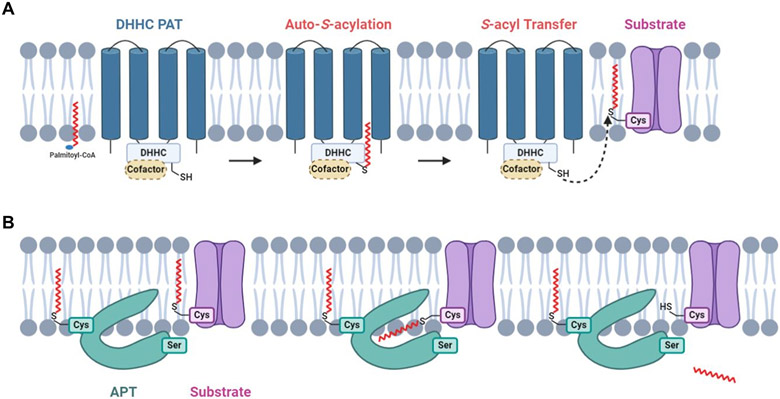
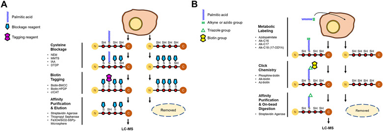
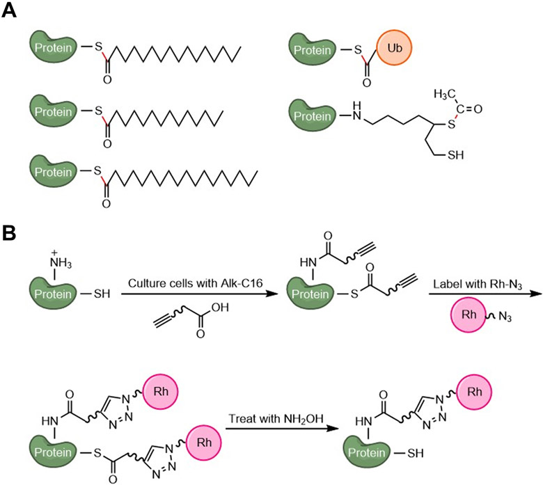
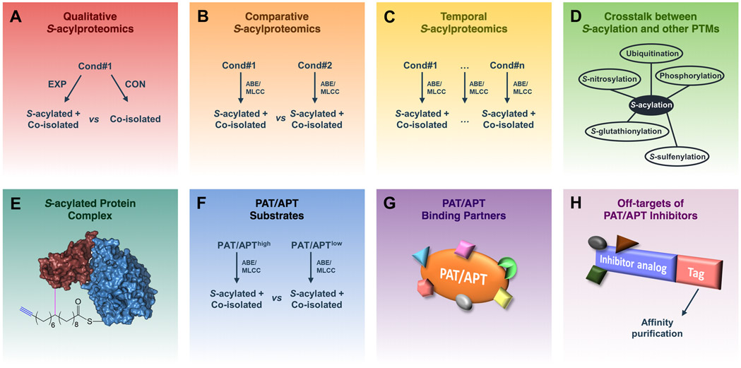
Protein S-acylation (commonly known as palmitoylation) is a widespread reversible lipid modification, which plays critical roles in regulating protein localization, activity, stability, and complex formation. The deregulation of protein S-acylation contributes to many diseases such as cancer and neurodegenerative disorders. The past decade has witnessed substantial progress in proteomic analysis of protein S-acylation, which significantly advanced our understanding of S-acylation biology. In this review, we summarized the techniques for the enrichment of S-acylated proteins or peptides, critically reviewed proteomic studies of protein S-acylation at eight different levels, and proposed major challenges for the S-acylproteomics field. In summary, proteome-scale analysis of protein S-acylation comes of age and will play increasingly important roles in discovering new disease mechanisms, biomarkers, and therapeutic targets.

Keywords: ABE; APT; MLCC; PAT; S-acylation; S-palmitoylation; click chemistry; palmitoylation; proteomics.


Figures

Figure 1.
Figure 1.. The biochemistry of protein S-acylation and de-S-acylation.
(A) DHHC-PATs are integral membrane proteins with active sites oriented toward the cytosol. A DHHC-PAT is first autoacylated on the DHHC cysteine residue, followed by a transfer of the S-acyl group to the acceptor cysteine residue of a substrate protein. Certain DHHC-PATs require a cofactor for proper functioning. (B) De-S-acylation enzymes can remove S-acyl groups from substrate S-acylproteins. A subpopulation of de-S-acylation enzymes are S-acylated and tethered to membrane, where their hydrophobic pockets capture S-acyl chains of substrate S-acylproteins, allowing enzymatic de-S-acylation.
Figure 2.
Figure 2.. Schematic of the methods for the enrichment of S-acylated proteins.
(A) In acyl-biotinyl exchange (ABE), free thiols on cysteines are blocked by an alkylation reagent (shown as a blue pentagon). S-acyl groups are specifically cleaved off by neutral hydroxylamine, and the newly exposed free thiols are labeled by a biotin-tag (shown as a purple double pentagon). The biotin-tagged (formerly S-acylated) proteins are captured by streptavidin affinity purification, eluted by a reducing agent, and analyzed by LC-MS/MS. (B) In metabolic labeling with a palmitic acid analog followed by click chemistry (MLCC), an alkyne- or azide (shown as a green triple bond)-functionalized palmitate analog is incorporated into S-acylation sites by the endogenous S-acylation machinery. After cell lysis, the palmitate analog-labeled (i.e., S-acylated) proteins are conjugated with a biotin analog (shown as a yellow double pentagon) by click chemistry (a triazole is shown as a green triangle), selectively enriched by streptavidin affinity purification, and digested by trypsin on beads.
Figure 3.
Figure 3.. Illustration of post-translational modifications that can be detected by ABE or MLCC.
(A) Representative thioester-linked PTMs that cannot be distinguished by ABE. Thioester bonds are shown in red. (B) Representative fatty acylation (S-acylation and N-acylation) that can be detected by MLCC. Hydroxylamine treatment enables the distinction between S-acylation and N-acylation.
Figure 4.
Figure 4.. Illustration of different types of proteomics applications in S-acylation studies.
(A) Qualitative S-acylproteomics focuses on profiling S-acylproteins and S-acylation sites as well as distinguishing them from co-isolated non-S-acylated forms. (B) Comparative S-acylproteomics compares the differences between S-acylproteomes under different conditions. (C) Temporal S-acylproteomics investigates the dynamic changes of protein S-acylation at different time points. (D) Proteomic analysis of the cross-talk between S-acylation and other PTMs. (E) Proteomic analysis of S-acylated protein complexes identifies proteins associated with the S-acylated form of a target protein. (F) Proteomic analysis of PAT/APT substrates by identifying proteins whose S-acylation levels are regulated by a specific PAT/APT. (G) Proteomic analysis of PAT/APT-binding partners. (H) Proteomic identification of off-targets of a PAT/APT inhibitor.
Figure 5.
Figure 5.. Stacked histogram of the numbers of publications from 2006 to 2020, which were grouped according to the study types.
(A) Stacked histogram in absolute numbers. (B) Stacked histogram in percentages.
Figure 6.
Figure 6.. Categorization of known human and mouse S-acylated proteins.
(A) Distribution of known human S-acylated proteins. (B) Distribution of known mouse S-acylated proteins. The numbers were retrieved from the SwissPalm database (v3).
Figure 7.
Figure 7.. Histograms of known substrates of human and mouse PATs/APTs.
(A) Histogram of known protein substrates of human PATs. (B) Histogram of known protein substrates of mouse PATs. (C) Histogram of known protein substrates of human APTs. (D) Histogram of known protein substrates of mouse APTs.
Figure 8.
Figure 8.. Histogram of protein interactors of human PATs/APTs.
The numbers were retrieved from the HuRI database (http://www.interactome-atlas.org/).

References

    1. Jiang H; Zhang X; Chen X; Aramsangtienchai P; Tong Z; Lin H Protein Lipidation: Occurrence, Mechanisms, Biological Functions, and Enabling Technologies. Chem. Rev 2018, 118 (3), 919–988. - PMC - PubMed
    1. Linder ME; Deschenes RJ Palmitoylation: Policing Protein Stability and Traffic. Nat. Rev. Mol. Cell Biol 2007, 8 (1), 74–84. - PubMed
    1. Muszbek L; Laposata M Covalent Modification of Platelet Proteins by Palmitate. Blood 1989, 74 (4), 1339–1347. - PubMed
    1. Muszbek L; Laposata M Myristoylation of Proteins in Platelets Occurs Predominantly through Thioester Linkages. J. Biol. Chem 1993, 268 (11), 8251–8255. - PubMed
    1. Muszbek L; Laposata M Covalent Modification of Proteins by Arachidonate and Eicosapentaenoate in Platelets. J. Biol. Chem 1993, 268 (24), 18243–18248. - PubMed

Publication types