

Temporal variations in the diagnostic performance of chest CT for Covid-19 depending on disease prevalence: Experience from North-Eastern France
- PMID: 33254065
- PMCID: PMC7836755
- DOI: 10.1016/j.ejrad.2020.109425
Temporal variations in the diagnostic performance of chest CT for Covid-19 depending on disease prevalence: Experience from North-Eastern France
Abstract
Rationale and objective: The purpose of this work was to analyze temporal variations in the diagnostic performance of chest CT for Covid-19 throughout the first wave, depending on disease prevalence variations between the ascending, peak and descending phases of the epidemic in North-Eastern France.
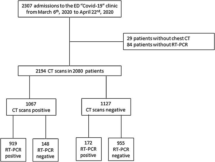
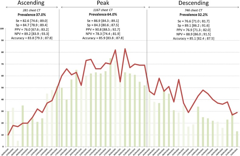
Materials and methods: From March 6th to April 22nd 2020, all consecutive adult patients referred to the "Covid-19 clinic" of our Emergency Department with the availability of chest CT and of at least one RT-PCR result were retrospectively included in the present study. Chest CT was considered positive when typical Covid-19 lesions were observed (bilateral and predominantly peripheral and sub-pleural ground glass opacities and/or alveolar consolidations). RT-PCR results were considered as the reference standard. Ascending, peak and descending phases were determined based on the number of CT scans performed daily. CT diagnostic performance were calculated and variations between phases were tested for equivalence or difference using Bayesian methods.
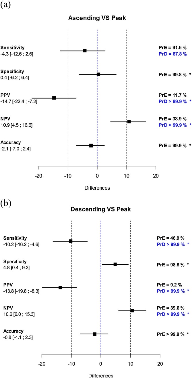
Results: 2194 consecutive chest CT were analyzed. Overall CT diagnostic performance was Se = 84.2 [82.0 ; 86.3], Sp = 86.6 [84.5 ; 88.5], PPV = 86.1 [84.0 ; 88.1], NPV = 84.7 [82.6 ; 86.7] and accuracy = 85.4 [83.9 ; 86.8], with no significant differences between chest and non-chest radiologists. Variations between the ascending (11 days, 281 chest CT, disease prevalence 37.0 %), the peak (18 days, 1167 chest CT, disease prevalence 64 %) and the descending phases (19 days, 746 chest CT, disease prevalence 32.2 %) were highest for PPV and NPV with a probability of difference >99.9 %, and smallest for accuracy and specificity with a probability of equivalence >98.8 %.
Conclusion: In a homogenous cohort of 2194 consecutive chest CT performed over a 7-week epidemic wave, we observed significant variations of CT predictive values whereas CT specificity appeared marginally affected.
Keywords: Covid-19; Diagnostic imaging; Lung diseases; Multidetector computed tomography; Reverse transcriptase polymerase chain reaction.
Copyright © 2020 Elsevier B.V. All rights reserved.
Conflict of interest statement
The author declares no sources of support or conflict of interest.
Figures
References
-
- Revel M.P., Parkar A.P., Prosch H., Silva M., Sverzellati N., Gleeson F., Brady A., R. European Society of, I. the European Society of Thoracic . European radiology; 2020. COVID-19 Patients and the Radiology Department - Advice From the European Society of Radiology (ESR) and the European Society of Thoracic Imaging (ESTI) - PMC - PubMed
MeSH terms
LinkOut - more resources
Full Text Sources
Other Literature Sources
Medical
Miscellaneous
