Review
. 2021 Feb 1:754:142363.
doi: 10.1016/j.scitotenv.2020.142363. Epub 2020 Sep 16.

Prospects of nanomaterials-enabled biosensors for COVID-19 detection

Affiliations
Review

Prospects of nanomaterials-enabled biosensors for COVID-19 detection

Manish Srivastava et al. Sci Total Environ. .

Abstract

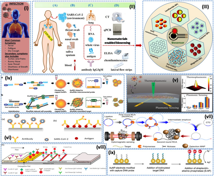
We are currently facing the COVID-19 pandemic which is the consequence of severe acute respiratory syndrome coronavirus (SARS-CoV-2). Since no specific vaccines or drugs have been developed till date for the treatment of SARS-CoV-2 infection, early diagnosis is essential to further combat this pandemic. In this context, the reliable, rapid, and low-cost technique for SARS-CoV-2 diagnosis is the foremost priority. At present reverse transcription polymerase chain reaction (RT-PCR) is the reference technique presently being used for the detection of SARS-CoV-2 infection. However, in a number of cases, false results have been noticed in COVID-19 diagnosis. To develop advanced techniques, researchers are continuously working and in the series of constant efforts, nanomaterials-enabled biosensing approaches can be a hope to offer novel techniques that may perhaps meet the current demand of fast and early diagnosis of COVID-19 cases. This paper provides an overview of the COVID-19 pandemic and nanomaterials-enabled biosensing approaches that have been recently reported for the diagnosis of SARS-CoV-2. Though limited studies on the development of nanomaterials enabled biosensing techniques for the diagnosis of SARS-CoV-2 have been reported, this review summarizes nanomaterials mediated improved biosensing strategies and the possible mechanisms that may be responsible for the diagnosis of the COVID-19 disease. It is reviewed that nanomaterials e.g. gold nanostructures, lanthanide-doped polysterene nanoparticles (NPs), graphene and iron oxide NPs can be potentially used to develop advanced techniques offered by colorimetric, amperometric, impedimetric, fluorescence, and optomagnetic based biosensing of SARS-CoV-2. Finally, critical issues that are likely to accelerate the development of nanomaterials-enabled biosensing for SARS-CoV-2 infection have been discussed in detail. This review may serve as a guide for the development of advanced techniques for nanomaterials enabled biosensing to fulfill the present demand of low-cost, rapid and early diagnosis of COVID-19 infection.

Keywords: Biosensors; COVID-19; Coronavirus; Nanomaterials; SARS-CoV-2.


Conflict of interest statement

Declaration of competing interest Authors of the manuscript declare there is no conflict of interests.

Figures

Unlabelled Image
Graphical abstract
Fig. 1
Fig. 1
Shows (A) 3D structure of SARS-CoV-2, (B) cross sectional representation of the viral structure with its proteins [modified background, adapted from: https://www.scientificanimations.com/coronavirus-symptoms-and-prevention-explained-through-medical-animation/] (https://en.wikipedia.org/wiki/Coronavirus, CC BY-SA 4.0) (C) Transmission electron microscope image of SARS-CoV-2. The virus is colorized in blue (adapted from the US Centers for Disease Control). [Adapted from: details on COVID-19; Public Health Image Library (PHIL), Centers for Disease Control and Prevention. https://phil.cdc.gov/Details.aspx?pid=23354 (accessed 2020/03/27).] (For interpretation of the references to colour in this figure legend, the reader is referred to the web version of this article.)
Fig. 2
Fig. 2
Schematic shows that one should take precautions to avoid infection through SARS-CoV-2 [credit: https://pixabay.com/; coronavirus 3D image adopted from https://www.scientificanimations.com/coronavirus-symptoms-and-prevention-explained-through-medical-animation/] (https://en.wikipedia.org/wiki/Coronavirus, CC BY-SA 4.0).
Fig. 3
Fig. 3
Nanomaterials-enabled biosensing approaches for respiratory viral infections (i) SARS-CoV-2 infection; revealing symptoms [modified from Mahapatra and Chandra, 2020] (ii) current diagnostic methods and portable biosensors for SARS-CoV-2 infection [modified from Cui and Zhou, 2020] (iii) different types of nanomaterials based biosensing for respiratory viral infections (iv) colorimetric detection of DNA based on disulfide induced self-assembly for MERS-CoV [Kim et al., 2019]. (v) dual-functional plasmonic photo-thermal biosensors for SARS-CoV-2 using AuNIs [Qiu et al., 2020]. (vi) optical fiber enabled biosensing [Nag et al., 2020], (vii) real-time optomagnetic detection of SARS-CoV-2 following homogeneous circle-to-circle amplification [Tian et al., 2020b], (viii) SARS-CoV-2 IgM-IgG combined antibody test [Li et al., 2020b, https://creativecommons.org/licenses/by/4.0/]. (ix) enzymatic electrochemical detection of SARS [Draz and Shafiee, 2018, Creative Commons Attribution (CC BY-NC) license, and also credit to report by Martínez-Paredes et al., 2009].
Fig. 4
Fig. 4
PPT enhancement in LSPR biosensing. (a) Schematic illustration of the hybridization of two complementary strands. (b) Real-time hybridization of RdRp-COVID and its cDNA sequence (RdRp-COVID-C) with or without the thermoplasmonic enhancement. (c) PPT enhancement on RdRp-COVID sequence detection at different concentrations. The error bars refer to the standard deviations of LSPR responses after reaching the steady conditions following the buffer flushing. (d) Schematic illustration of inhibited hybridization of two partially matched sequences. The red arrows indicated the mismatch bases of RdRp-SARS and functionalized cDNA of RdRp-COVID. (e) Discrimination of two similar sequences with PPT heat. The laser was applied at 200 s and switched off at 700 s. (f) RdRp-SARS sequence dissociation from the immobilized RdRp-COVID-C sequence. The original phase responses (red dots) and the corresponding smoothed means (black curve) are shown. [Reproduced with permission from: Qiu et al., 2020]. (For interpretation of the references to colour in this figure legend, the reader is referred to the web version of this article.)
Fig. 5
Fig. 5
Schematic representation for the Selective Naked-Eye Detection of SARS-CoV-2 RNA Mediated by the Suitably Designed ASO-Capped AuNPs [reproduced with permission from: Moitra et al., 2020].
Fig. 6
Fig. 6
Design and fabrication of the developed LFIA assay. (A) Lateral flow test strip. (B) Assay. [Reproduced with permission from: Chen et al., 2020].
Fig. 7
Fig. 7
Detection of SARS-CoV-2 virus from clinical samples. (A) Schematic diagram for the COVID-19 FET sensor for detection of SARS-CoV-2 virus from COVID-19 patients. (B, C) Comparison of response signal between normal samples and patient ones. (D) Real-time response of COVID-19 FET towards SARS-CoV-2 clinical sample and (C) related dose-dependent response curve. [Reproduced with permission from: Seo et al., 2020].

References

    1. Alifano M., Alifano P., Forgez P., Iannelli A. Renin-angiotensin system at the heart of COVID-19 pandemic. Biochimie. 2020;174:30–33. doi: 10.1016/j.biochi.2020.04.008. - DOI - PMC - PubMed
    1. Augustine S., Singh J., Srivastava M., Sharma M., Das A., Malhotra B.D. Recent advances in carbon based nanosystems for cancer theranostics. [10.1039/C7BM00008A] Biomaterials Science. 2017;5(5):901–952. doi: 10.1039/c7bm00008a. - DOI - PubMed
    1. Bai Y., Yao L., Wei T., Tian F., Jin D.-Y., Chen L., Wang M. Presumed asymptomatic carrier transmission of COVID-19. JAMA. 2020;323(14):1406–1407. doi: 10.1001/jama.2020.2565. - DOI - PMC - PubMed
    1. Banerjee R., Jaiswal A. Recent advances in nanoparticle-based lateral flow immunoassay as a point-of-care diagnostic tool for infectious agents and diseases. [10.1039/C8AN00307F] Analyst. 2018;143(9):1970–1996. doi: 10.1039/c8an00307f. - DOI - PubMed
    1. Barnett J.M., Monnier B.M., Tyler S., West D., Ballantine-Dykes H., Regan E.…Luxton R. Initial trail results of a magnetic biosensor for the rapid detection of Porcine Reproductive and Respiratory Virus (PRRSV) infection. Sensing and Bio-Sensing Research. 2020;27 doi: 10.1016/j.sbsr.2019.100315. - DOI