. 2020 Nov 24;9(12):2530.
doi: 10.3390/cells9122530.

Smart Nanofibers with Natural Extracts Prevent Senescence Patterning in a Dynamic Cell Culture Model of Human Skin

Affiliations

Smart Nanofibers with Natural Extracts Prevent Senescence Patterning in a Dynamic Cell Culture Model of Human Skin

Emanuela Bellu et al. Cells. .

Erratum in

Abstract

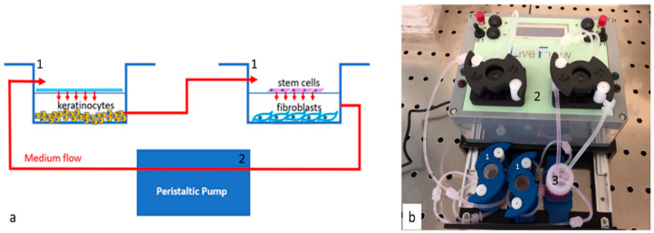
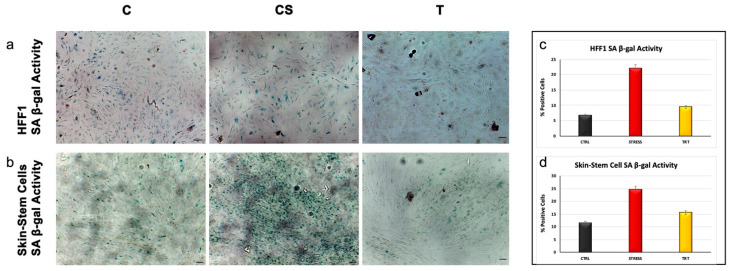
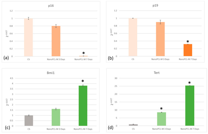
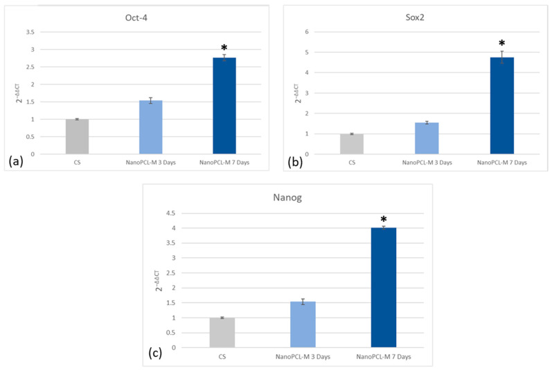
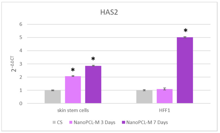
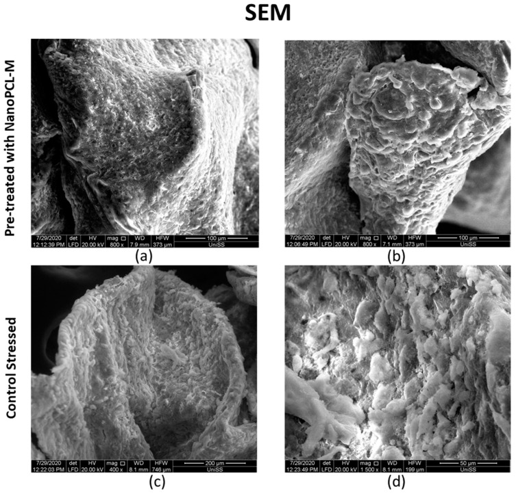
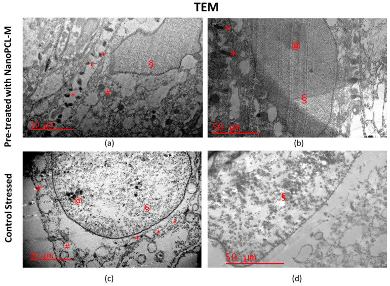
Natural cosmetic products have recently re-emerged as a novel tool able to counteract skin aging and skin related damages. In addition, recently achieved progress in nanomedicine opens a novel approach yielding from combination of modern nanotechnology with traditional treatment for innovative pharmacotherapeutics. In the present study, we investigated the antiaging effect of a pretreatment with Myrtus communis natural extract combined with a polycaprolactone nanofibrous scaffold (NanoPCL-M) on skin cell populations exposed to UV. We set up a novel model of skin on a bioreactor mimicking a crosstalk between keratinocytes, stem cells and fibroblasts, as in skin. Beta-galactosidase assay, indicating the amount of senescent cells, and viability assay, revealed that fibroblasts and stem cells pretreated with NanoPCL-M and then exposed to UV are superimposable to control cells, untreated and unexposed to UV damage. On the other hand, cells only exposed to UV stress, without NanoPCL-M pretreatment, exhibited a significantly higher yield of senescent elements. Keratinocyte-based 3D structures appeared disjointed after UV-stress, as compared to NanoPCL-M pretreated samples. Gene expression analysis performed on different senescence associated genes, revealed the activation of a molecular program of rejuvenation in stem cells pretreated with NanoPCL-M and then exposed to UV. Altogether, our results highlight a future translational application of NanoPCL-M to prevent skin aging.

Keywords: 4D dynamic model; biophysics; cell senescence; cellular mechanisms; nanofibers; natural extracts; precision medicine; skin aging; stem cells.


Conflict of interest statement

The authors declare no conflict of interest. The funders had no role in the design of the study; in the collection, analyses, or interpretation of data; in the writing of the manuscript, or in the decision to publish the results.

Figures

Figure 1
Figure 1
Scheme (a) and picture (b) of bioreactor. Panel a represent the chambers (Live Box2; blue lines (1)) and the cells cultured in every chamber. The chamber and the pump (Live Flow (2)) connections are visible by medium flow (red lines and arrows in the scheme). Panel b is the picture of the bioreactor used (IVTech) the chambers Live Box2 are blue (1); number (2) indicates the pump and number (3) the reservoir for the culture medium.
Figure 2
Figure 2
Images acquired with SEM microscope showing nanofiber structure. Panel (a) show nanofiber at 15 k magnification. Panel (b) show nanofiber at 3 k magnification.
Figure 3
Figure 3
Release of myrtle extracts from NanoPCL-M for seven days. The amount of extracts is evaluated as absorbance OD detected at 405 nm, and expressed as mg/day. Error bars indicated standard deviation of the experiments (n = 6).
Figure 4
Figure 4
(a) The effect of NanoPCL-M on HFF1 fibroblasts and skin stem cells after three and seven days of pretreatment and then UV exposure. Graphs show the percentage of cellular metabolic activity as compared to negative control C (100%). CS represent cells untreated with NanoPCL-M and exposed to UV (stressed control), and C represents negative control. (b) effect of nanofiber in PCL (poly ε-caprolactone) (CTRL PCL) on stem cells compared with cells untreated (CTRL). Error bars represent standard deviation. * p value ≤ 0.05
Figure 5
Figure 5
Senescence-associated β-galactosidase activity evaluated in HFF1 (a) and skin stem cells (b) after seven days. Cells pretreated with NanoPCL-M (T) are compared to control untreated cells (C) and UV stress control (CS). Scale bar = 100 µm. The number of blue positive HFF1 (c) and skin stem (d) was calculated using ImageJ. Data are expressed as mean ± SD.
Figure 6
Figure 6
Effect of NanoPCL-M pretreatment on the expression of p16 (a), p19 (b), Bmi1 (c) and TERT (d). Skin stem cells exposed (T) or not (CS) to three to seven days of NanoPCL-M pretreatment were stressed by UV light. The amount of mRNA from NanoPCL-M treated cells was normalized to HPRT1 and was plotted as fold change (2−∆∆CT) relative to the mRNA expression of UV stress control (CS). * p value ≤ 0.05.
Figure 7
Figure 7
Effect of NanoPCL-M pretreatment on the expression of Oct-4 (a), Sox2 (b) and Nanog (c). Skin stem cells exposed (T) or not (CS) to three to seven days of NanoPCL-M pretreatment were stressed by UV light. The amount of mRNA from NanoPCL-M treated cells was normalized to HPRT1 and was plotted as fold change (2−∆∆CT) relative to the mRNA expression of UV stress control (CS). * p value ≤ 0.05
Figure 8
Figure 8
Effect of NanoPCL-M pretreatment on the expression of HAS2. Skin stem cells and HFF1 exposed (T) or not (CS) to three to seven days of NanoPCL-M pretreatment were stressed by UV light. The amount of mRNA from NanoPCL-M treated cells was normalized to HPRT1 and was plotted as fold change (2−∆∆CT) relative to the mRNA expression of UV stress control (CS). * p value ≤ 0.05
Figure 9
Figure 9
Images acquired with SEM microscope showing keratinocytes 3D organization. (a,b) represent UV stressed samples pretreated (T) with NanoPCL-M for seven days. (c,d) represent UV stressed samples (CS) kept in culture for seven days without any pretreatment. Scale bar is indicated in each panel.
Figure 10
Figure 10
Images acquired with TEM showing keratinocytes 3D organization. Scale bar = 50 um. (a,b) represent UV stressed samples pretreated (T) with NanoPCL-M for seven days. (c), (d) represent UV stressed samples (CS) kept in culture for seven days without any pretreatment. * Desmosomes; § Chromatin; # Vacuoles. Desmosome junctions (*) appeared regular and preserved intracellular junctions in T samples ((a,b)*); on the contrary they were altered and degenerated in morphology in CS samples (panel c *). Chromatin (§) was homogenous like euchromatin in the T sample ((a,b) *) with visible nucleolus ((b) @). On the other hand chromatin (§) of CS samples appeared disorganized, not homogenous ((c,d) §) and fragmented ((d) §). Vacuoles shows irregular membrane in CS samples ((c) #) and are present in low number and smaller in T samples ((a) #).

References

    1. Kim E.K., Kim H.O., Park Y.M., Park C.J., Yu D.S., Lee J.Y. Prevalence and risk factors of depression in geriatric patients with dermatological diseases. Ann. Dermatol. 2013;25:278–284. doi: 10.5021/ad.2013.25.3.278. - DOI - PMC - PubMed
    1. Draelos Z.D. Cosmetics and skin care products. A historical perspective. Dermatol. Clin. 2000;18:557–559. doi: 10.1016/S0733-8635(05)70206-0. - DOI - PubMed
    1. Tundis R., Loizzo M.R., Bonesi M., Menichini F. Potential role of natural compounds against skin aging. Curr. Med. Chem. 2015;22:1515–1538. doi: 10.2174/0929867322666150227151809. - DOI - PubMed
    1. Barry B.W. Novel mechanisms and devices to enable successful transdermal drug delivery. Eur. J. Pharm. Sci. 2001;14:101–114. doi: 10.1016/S0928-0987(01)00167-1. - DOI - PubMed
    1. Farokhzad O.C., Langer R. Impact of nanotechnology on drug delivery. ACS Nano. 2009;3:16–20. doi: 10.1021/nn900002m. - DOI - PubMed

Publication types

LinkOut - more resources