. 2020 Nov 24;12(12):3491.
doi: 10.3390/cancers12123491.

Sensitization of Cutaneous Primary Afferents in Bone Cancer Revealed by In Vivo Calcium Imaging

Affiliations

Sensitization of Cutaneous Primary Afferents in Bone Cancer Revealed by In Vivo Calcium Imaging

Larissa de Clauser et al. Cancers (Basel). .

Abstract

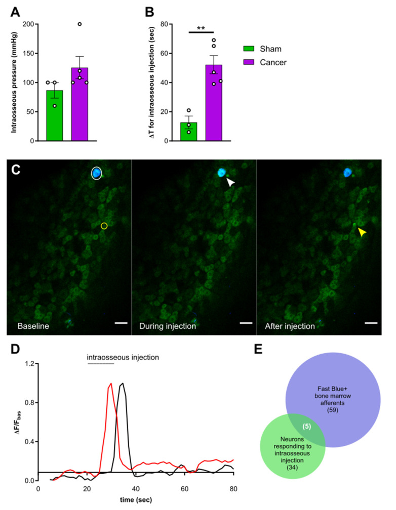
Cancer-induced bone pain (CIBP) is a complex condition, comprising components of inflammatory and neuropathic processes, but changes in the physiological response profiles of bone-innervating and cutaneous afferents remain poorly understood. We used a combination of retrograde labelling and in vivo calcium imaging of bone marrow-innervating dorsal root ganglia (DRG) neurons to determine the contribution of these cells in the maintenance of CIBP. We found a majority of femoral bone afferent cell bodies in L3 dorsal root ganglia (DRG) that also express the sodium channel subtype Nav1.8-a marker of nociceptive neurons-and lack expression of parvalbumin-a marker for proprioceptive primary afferents. Surprisingly, the response properties of bone marrow afferents to both increased intraosseous pressure and acid were unchanged by the presence of cancer. On the other hand, we found increased excitability and polymodality of cutaneous afferents innervating the ipsilateral paw in cancer bearing animals, as well as a behavioural phenotype that suggests changes at the level of the DRG contribute to secondary hypersensitivity. This study demonstrates that cutaneous afferents at distant sites from the tumour bearing tissue contribute to mechanical hypersensitivity, highlighting these cells as targets for analgesia.

Keywords: CIBP; DRG; bone afferents; in vivo imaging; nociception; peripheral sensitization; secondary hypersensitivity.


Conflict of interest statement

The authors declare no conflict of interest. The funders had no role in the design of the study; in the collection, analyses, or interpretation of data; in the writing of the manuscript, or in the decision to publish the results.

Figures

Figure 1
Figure 1
Bone marrow afferents are medium-to large sized neurons expressing the nociceptive marker Nav1.8 and lacking expression of proprioceptive marker parvalbumin. (A) Rostrocaudal distribution of all Fast Blue+ retrogradely labelled bone marrow afferents within the analysed ipsilateral lumbar dorsal root ganglia (DRG) (L2–L5). (B) Proportion of Fast Blue+ neurons within the Nav1.8-cre expressing population of DRG neurons throughout lumbar L2–L5. (C) Proportion of bone marrow afferents based on soma size. (DE) Representative images of lumbar 3 DRG immunostained for TrkA (green) and Fast Blue retrogradely traced (blue) femoral bone marrow afferents in combination with tdTomato expressing neurons (red) driven by (D) Nav1.8-cre, (E) Tmem233-cre, and (F) Pvalb-cre. White arrows indicate double positive Fast Blue/TrkA neurons; yellow arrows indicate triple positive neurons. Scale bar = 100 µm.
Figure 2
Figure 2
Pain behaviour in Pirt-GCaMP3-expressing mice with cancer-induced bone pain (CIBP). (A) Survival curve after surgery for sham (green line, n = 22) and cancer animals (purple line, n = 17) with endpoint defined as clear limping on the affected limb. Black bars indicate individual dropouts. (B) Ongoing pain was measured by percentage weight bearing on the affected limb. (C) Mechanical withdrawal threshold to von Frey filaments in cancer (n = 12) and sham animals (n = 21). (D) Number of nocifensive responses (guarding, licking, flinching) observed during the 2 min period after palpation of the distal femur head in cancer bearing (n = 5) and sham (n = 11) mice. (E) Mechanical withdrawal thresholds to application of the Randall–Selitto apparatus to the paw in cancer (n = 6) and sham animals (n = 7). (F). Thermosensation measured by response threshold to 50 °C hot-plate in cancer (n = 6) and sham mice (n = 4). * p < 0.05; ** p < 0.01; *** p < 0.001.
Figure 3
Figure 3
Response properties of cutaneous afferents of the hindpaw. (A) Percentage of total neurons responding to pinch, heat, and cold in cancer (purple, n = 10) and sham animals (green, n = 15). (B) Percentage of polymodal cutaneous afferents in L3 DRG of cancer and sham mice. (C) Percentage of coupled responses over total responses of the same stimulus modality for pinch and heat in sham and cancer bearing mice. (D,E) Response intensity of mechanosensitive (D) and heat sensitive (E) cutaneous afferents expressed as maximum fluorescence intensity during stimulus application vs. baseline. (F,G) Size distribution of mechanosensitive (n = 147 neurons in cancer, n = 170 neurons in sham) (F) and heat sensitive (n = 147 in cancer, n = 366 in sham) cutaneous afferents (G). * p < 0.05; ** p < 0.01; *** p < 0.001, **** p < 0.0001
Figure 4
Figure 4
Experimental setup for in vivo calcium imaging of cutaneous and bone marrow afferents. Adult mice expressing GCaMP3 under the control of the promoter Pirt were injected with Lewis Lung carcinoma (LLC) cells or vehicle and Fast Blue retrograde tracer into the distal femur head. (1) To measure secondary hypersensitivity, noxious pinch, hot water (55 °C), and ice-cold water (0 °C) were applied to the plantar surface of the affected paw. (2) To activate bone afferents 10 µL solution were delivered to the femoral marrow through a syringe and pressure was recorded. (3) Step 1 was repeated to determine if activation of bone afferents resulted in changes in excitability of distal cutaneous afferents.
Figure 5
Figure 5
Activation of femoral bone marrow afferents through intraosseous injection. (A) Intraosseous injection pressure at start of solution flow in cancer (purple, n = 5) and sham animals (green, n = 3). (B) Time elapsed from start of solution flow until delivery of 10 µL into the femoral bone marrow. (C) Representative images taken from a recording and showing the response of one Fast Blue labelled bone afferent (white arrow) responding during intraosseous injection and one unlabelled responding after (yellow arrow). Scale bar = 50 µm. (D) Example traces showing ΔF/Fbasal of bone afferents responding during (red trace) or shortly after (black trace) injection into the mouse bone marrow. Both types of responders were included in the analysis. (E) Venn diagram showing the overlap between Fast Blue+ cells (blue) and neurons responding to bone marrow injection (green) (data from animals with specific Fast Blue labelling, n = 6). ** p < 0.01.
Figure 6
Figure 6
Response properties of femoral bone marrow afferents. (A) Percentage of bone afferents on total number of neurons responding to plantar stimulation in sham (green) and cancer animals (purple) receiving intraosseous injection of saline (solid fill, n = 7 for sham, n = 5 for cancer) or acid (pattern fill, n = 7 for sham, n = 4 for cancer). (B) Maximum fluorescence intensity of bone marrow afferents responding to intraosseous injection (number of neurons: n = 14 for sham saline, n = 14 for cancer saline, n = 18 for sham acid, n = 8 for cancer acid). (C) Size distribution of bone afferents responding to saline (full line) or acid (stitched line) in cancer and sham animals. (D) Percentage of responding cutaneous afferents before (solid fill boxes, pinch = grey, heat = red, blue = cold) and after (pattern fill boxes) stimulation of bone marrow afferents.

Similar articles

Cited by

References

    1. Van Den Beuken-Van Everdingen M.H.J., Hochstenbach L.M.J., Joosten E.A.J., Tjan-Heijnen V.C.G., Janssen D.J.A. Update on Prevalence of Pain in Patients with Cancer: Systematic Review and Meta-Analysis. J. Pain Symptom Manag. 2016;51:1070–1090. doi: 10.1016/j.jpainsymman.2015.12.340. - DOI - PubMed
    1. Coleman R.E. Metastatic bone disease: Clinical features, pathophysiology and treatment strategies. Cancer Treat. Rev. 2001;27:165–176. doi: 10.1053/ctrv.2000.0210. - DOI - PubMed
    1. Coleman R.E. Clinical features of metastatic bone disease and risk of skeletal morbidity. Clin. Cancer Res. 2006;12:6243–6250. doi: 10.1158/1078-0432.CCR-06-0931. - DOI - PubMed
    1. Grond S., Zech D., Diefenbach C., Radbruch L., Lehmann K.A. Assessment of cancer pain: A prospective evaluation in 2266 cancer patients referred to a pain service. Pain. 1996;64:107–114. doi: 10.1016/0304-3959(95)00076-3. - DOI - PubMed
    1. Scarpi E., Calistri D., Klepstad P., Kaasa S., Skorpen F., Habberstad R., Nanni O., Amadori D., Maltoni M. Clinical and Genetic Factors Related to Cancer-Induced Bone Pain and Bone Pain Relief. Oncologist. 2014;19:1276–1283. doi: 10.1634/theoncologist.2014-0174. - DOI - PMC - PubMed

LinkOut - more resources