. 2020 Nov 24;13(23):5311.
doi: 10.3390/ma13235311.

Behaviour of Human Oral Epithelial Cells Grown on Invisalign® SmartTrack® Material

Affiliations

Behaviour of Human Oral Epithelial Cells Grown on Invisalign® SmartTrack® Material

Michael Nemec et al. Materials (Basel). .

Abstract

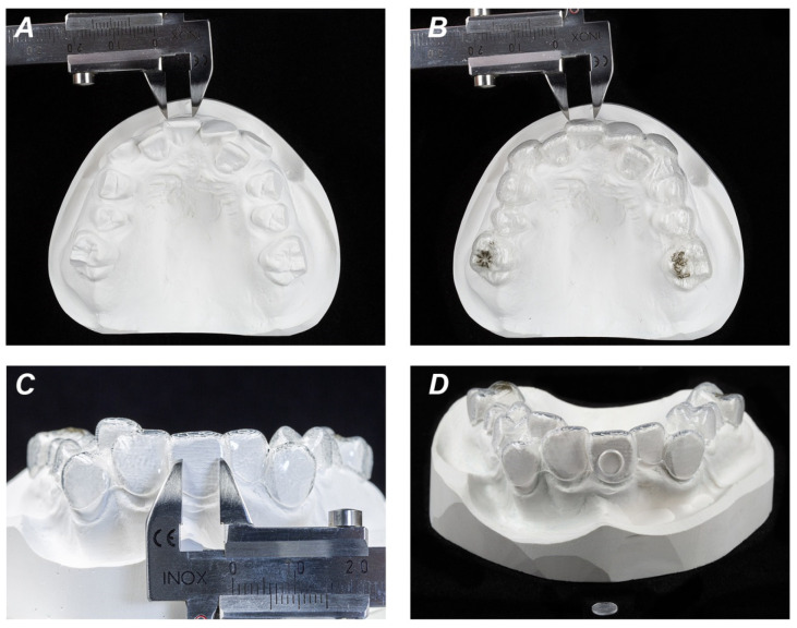
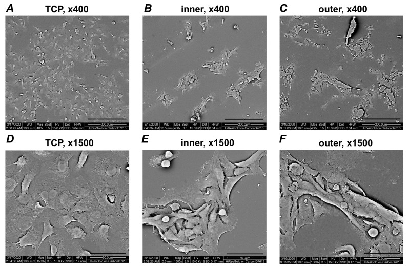
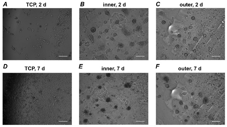
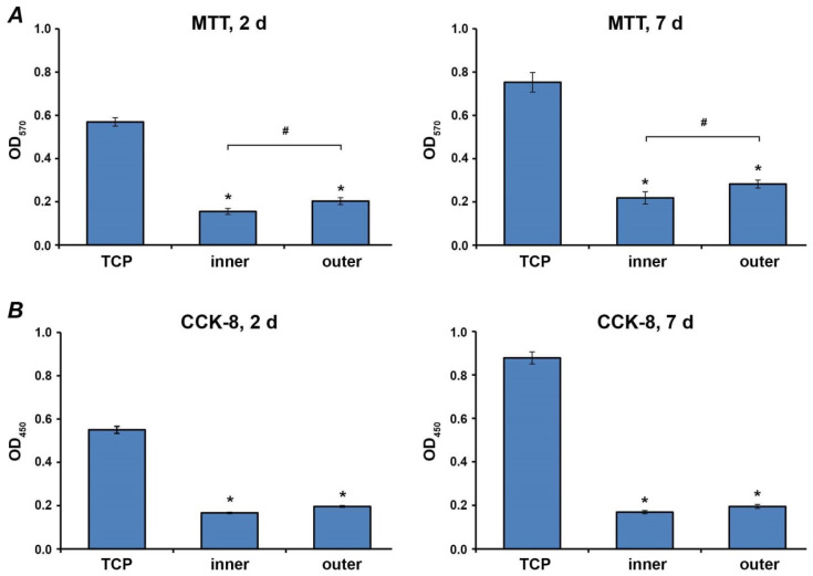
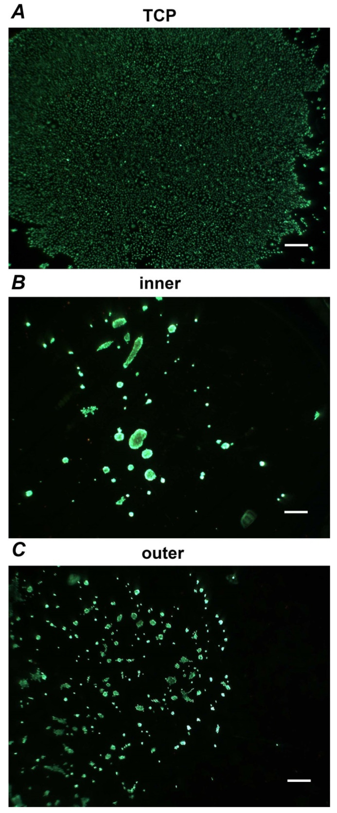
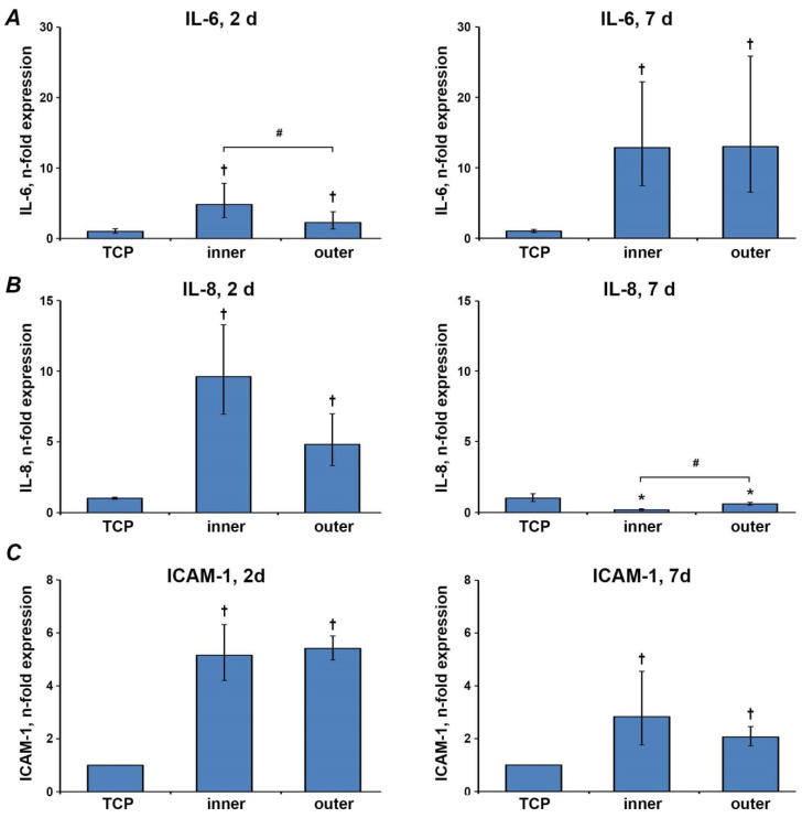
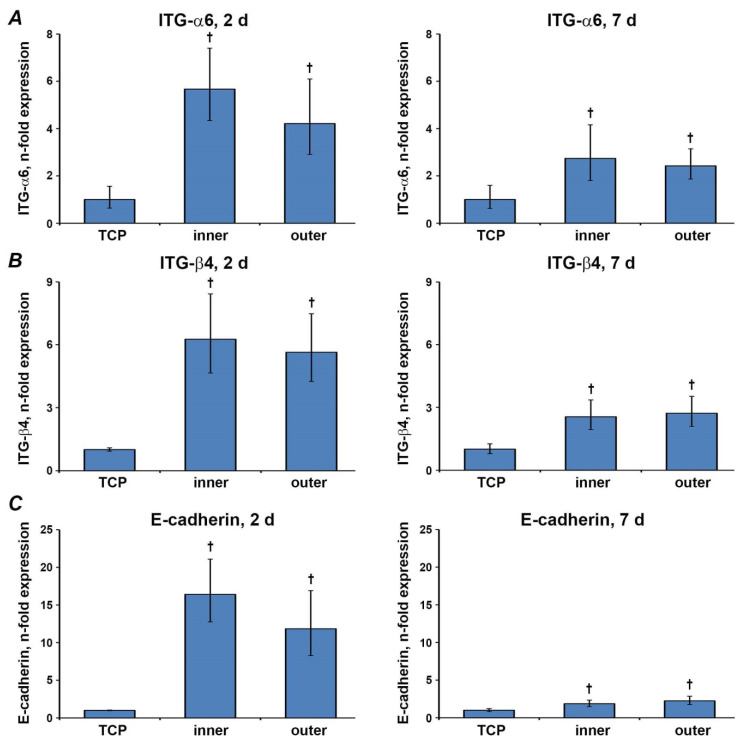
Invisalign aligners have been widely used to correct malocclusions, but their effect on oral cells is poorly known. Previous research evaluated the impact of aligners' eluates on various cells, but the cell behavior in direct contact with aligners is not yet studied. In the present study, we seeded oral epithelial cells (cell line Ca9-22) directly on Invisalign SmartTrack material. This material is composed of polyurethane and co-polyester and exhibit better mechanical characteristics compared to the predecessor. Cell morphology and behavior were investigated by scanning electron microscopy and an optical cell moves analyzer. The effect of aligners on cell proliferation/viability was assessed by cell-counting kit (CCK)-8 and 3,4,5-dimethylthiazol-2-yl-2,5-diphenyl tetrazolium bromide (MTT) assay and live/dead staining. The expression of inflammatory markers and proteins involved in epithelial barrier function was measured by qPCR. Cells formed cluster-like structures on aligners. The proliferation/viability of cells growing on aligners was significantly lower (p < 0.05) compared to those growing on tissue culture plastic (TCP). Live/dead staining revealed a rare occurrence of dead cells on aligners. The gene expression level of all inflammatory markers in cells grown on aligners' surfaces was significantly increased (p < 0.05) compared to cells grown on TCP after two days. Gene expression levels of the proteins involved in barrier function significantly increased (p < 0.05) on aligners' surfaces after two and seven days of culture. Aligners' material exhibits no cytotoxic effect on oral epithelial cells, but alters their behavior and the expression of proteins involved in the inflammatory response, and barrier function. The clinical relevance of these effects has still to be established.

Keywords: Invisalign; SmartTrack; aligner; epithelial barrier; in vitro; inflammation; oral epithelial cells; orthodontics; proliferation.


Conflict of interest statement

All authors declare no conflict of interest.

Figures

Figure 1
Figure 1
Preparation of orthodontic aligners used in the study. Conventional orthodontic plaster models were ground plane in the region of buccal areas. Required diameter of 6 mm of flat surface was confirmed by a conventional orthodontic caliper (A). Flat aligner surface of 6 mm equivalent to buccal plaster cast surface (B,C). Discs of 6 mm in diameter were cut out of regions with flat crown surfaces and used in cell culture experiments (D).
Figure 2
Figure 2
Scanning electron microscopy analysis of Ca9-22 cells grown on different surfaces. Pictures were taken at two different magnifications (upper row, ×400; lower row, ×1500). Cells seeded on TCP (control; A,D), inner (B,E) and outer (C,F) aligners’ surfaces are shown. Pictures were taken after seven days of culture. Scale bars correspond to 50 µm (AC) or 200 µm (DF).
Figure 3
Figure 3
Live cell analyzer pictures. Ca9-22 cells were grown on different surfaces and the pictures were taken every 30 min. Cells seeded on TCP (control; A,D), inner (B,E) and outer (C,F) aligners’ surfaces are shown. Shown pictures were taken after two (AC) and seven days (DF) of culture. Scale bars correspond to 200 µm.
Figure 4
Figure 4
Proliferation/viability of Ca9-22 cells grown on different surfaces. Ca9-22 cells were cultured on TCP, inner and outer aligners’ surfaces. Proliferation/viability was measured after two and seven days incubation by MTT (A) and CCK-8 (B) assays. Cells grown on tissue culture plastic served as control. Y-axis represents the optical density (OD) measured at 570 nm and 450 nm. Data represents mean ± s.e.m. of four independent experiments. *—significantly lower compared to control; #—significantly different between groups. p < 0.05.
Figure 5
Figure 5
Live/dead Staining. Ca9-22 cells were grown on TCP (A), inner (B) and outer (C) aligners’ surfaces for seven days and stained with Live/dead staining kit. Vital cells are visible as green while the dead cells are presented red. Images are taken from representative experiment. Scale bar correspond to 500 µm.
Figure 6
Figure 6
Gene expression of inflammatory markers. Gene expression of IL-6 (A), IL-8 (B) and ICAM1 (C) after two and seven days in Ca9-22 cells grown on different aligners’ surfaces were measured by qPCR. Y-axes represent n-fold expression in relation to Ca9-22 cells grown on TCP (n-fold expression = 1), calculated using 2−ΔΔCt method. Data are presented as the mean ± s.e.m. of four independent experiments. †—significantly higher compared to control; *—significantly lower compared to control; #—significantly different between groups. p < 0.05.
Figure 7
Figure 7
Gene expression of epithelial barrier markers. Gene expression of ITGα-6 (A), ITGβ-4 (B) and E-cadherin(C) after two and seven days in Ca9-22 cells grown on different aligners’ surfaces and TCP were measured by qPCR. Y-axes represent n-fold expression in relation to Ca9-22 cells grown on TCP (n-fold expression = 1), calculated using 2−ΔΔCt method. Data are presented as the mean ± s.e.m. of four independent experiments. † Significantly higher compared to control; p < 0.05.

References

    1. Rossini G., Parrini S., Castroflorio T., Deregibus A., Debernardi C.L. Efficacy of clear aligners in controlling orthodontic tooth movement: A systematic review. Angle Orthod. 2015;85:881–889. doi: 10.2319/061614-436.1. - DOI - PMC - PubMed
    1. Rosvall M.D., Fields H.W., Ziuchkovski J., Rosenstiel S.F., Johnston W.M. Attractiveness, acceptability, and value of orthodontic appliances. Am. J. Orthod. Dentofac. Orthop. 2009;135:276.e1–276.e12. doi: 10.1016/j.ajodo.2008.07.011. - DOI - PubMed
    1. Bernard G., Rompre P., Tavares J.R., Montpetit A. Colorimetric and spectrophotometric measurements of orthodontic thermoplastic aligners exposed to various staining sources and cleaning methods. Head Face Med. 2020;16:2. doi: 10.1186/s13005-020-00218-2. - DOI - PMC - PubMed
    1. Katchooi M., Cohanim B., Tai S., Bayirli B., Spiekerman C., Huang G. Effect of supplemental vibration on orthodontic treatment with aligners: A randomized trial. Am. J. Orthod. Dentofac. Orthop. 2018;153:336–346. doi: 10.1016/j.ajodo.2017.10.017. - DOI - PubMed
    1. Condo R., Pazzini L., Cerroni L., Pasquantonio G., Lagana G., Pecora A., Mussi V., Rinaldi A., Mecheri B., Licoccia S., et al. Mechanical properties of “two generations” of teeth aligners: Change analysis during oral permanence. Dent. Mater. J. 2018;37:835–842. doi: 10.4012/dmj.2017-323. - DOI - PubMed

LinkOut - more resources