. 2020 Nov 24;8(12):531.
doi: 10.3390/biomedicines8120531.

TrkB-Targeted Therapy for Mucoepidermoid Carcinoma

Affiliations

TrkB-Targeted Therapy for Mucoepidermoid Carcinoma

Vivian P Wagner et al. Biomedicines. .

Abstract

The brain-derived neurotrophic factor (BDNF)/tyrosine receptor kinase B (TrkB) pathway was previously associated with key oncogenic outcomes in a number of adenocarcinomas. The aim of our study was to determine the role of this pathway in mucoepidermoid carcinoma (MEC). Three MEC cell lines (UM-HMC-2, H253 and H292) were exposed to Cisplatin, the TrkB inhibitor, ANA-12 and a combination of these drugs. Ultrastructural changes were assessed through transmission electron microscopy; scratch and Transwell assays were used to assess migration and invasion; and a clonogenic assay and spheroid-forming assay allowed assessment of survival and percentage of cancer stem cells (CSC). Changes in cell ultrastructure demonstrated Cisplatin cytotoxicity, while the effects of ANA-12 were less pronounced. Both drugs, used individually and in combination, delayed MEC cell migration, invasion and survival. ANA-12 significantly reduced the number of CSC, but the Cisplatin effect was greater, almost eliminating this cell population in all MEC cell lines. Interestingly, the spheroid forming capacity recovered, following the combination therapy, as compared to Cisplatin alone. Our studies allowed us to conclude that the TrkB inhibition, efficiently impaired MEC cell migration, invasion and survival in vitro, however, the decrease in CSC number, following the combined treatment of ANA-12 and Cisplatin, was less than that seen with Cisplatin alone; this represents a limiting factor.

Keywords: adenocarcinoma; cell biology; head and neck neoplasms; salivary gland neoplasms; therapeutics.


Conflict of interest statement

The authors declare no conflict of interest.

Figures

Figure 1
Figure 1
(A) Representative images of BDNF and pTrkB immunostaining in normal minor (MiSG) and major salivary glands (MaSG) and mucoepidermoid carcinoma (MEC) with different grades. Note the absence of pTrkB expression in mucous cells (arrow) and the presence of expression in cells associated with perineural invasion (asterisk). (B) The percentage of BDNF- and pTrkB-positive cells was significantly higher in MEC, compared to normal salivary gland tissue (scores obtained from the joint analysis of MaSG and MiSG) (p < 0.0001 and p = 0.0075, respectively, Mann-Whitney Test). (C) No significant difference was detected among the different MEC grades concerning BDNF and pTrkB expression (p > 0.05, Kruskal-Wallis Test), however, high-grade tumours presented a tendency for elevated percentage of pTrkB positive cells. (D) BDNF and pTrkB transcript levels in normal salivary gland primary cells (NSGPC) and MEC cell lines revealed higher BDNF expression in all MEC cell lines compared to NSGPC. Interestingly, high TrkB expression was observed in the high-grade salivary gland MEC (H253) with lower expression in the pulmonary MEC cell line (H292). The levels of UM-HMC-2 were similar to NSGPC (** p < 0.01; **** p < 0.0001, and NS p > 0.05).
Figure 2
Figure 2
Representative images of MEC cells ultrastructural morphology following ANA-12 and cisplatin administration. Note, Cisplatin induced a high number of apoptotic changes such as decrease in cells size, “cup-shaped” chromatin condensation, convolution of the nuclear and cellular outlines, loss of cell contact, loss of specialized surface elements such as microvilli and cell–cell junctions, and ultimately cell fragmentation and formation of apoptotic bodies. Cytosolic constituents released into the extracellular space can be observed indicating a process of secondary necrosis. Scale bar—2 µm.
Figure 3
Figure 3
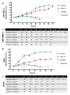
(A) UM-HMC-2 wound closure following ANA-12, Cisplatin and combined treatment. Note that the combined treatment presented the most promising results to delay wound closure in this cell line. (B) H253 wound closure following ANA-12, Cisplatin and combined treatment. Note that Cisplatin and the combined treatment results were similar and ANA-12 effects occurred earlier and in a greater magnitude, compared to UM-HMC-2 (* p < 0.05; ** p < 0.01; *** p < 0.001; **** p < 0.0001 and NS p > 0.05).
Figure 4
Figure 4
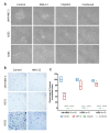
(A) Representative images of the invading cells following ANA-12, Cisplatin and combined treatment. Time of invasion is presented on the right side. (B) Quantitative analysis revealed that all treatments were effective in disrupting cell invasive capacities. Yet, note that Cisplatin and combined treatment achieved the most promising results for all cell lines (**** p < 0.0001).
Figure 5
Figure 5
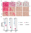
(A) Representative images of phase contrast microscopy during the clonogenic assay. Note that under Cisplatin and combined treatment, MEC cells remained attached and with a healthy morphology, but did not present the capacity to form colonies. (B) Comparison of stained colonies in control and ANA-12 groups. Note that the number and size of colonies is reduced following TrkB inhibition. (C) Quantitative analysis revealed a significant reduction in the number of surviving colonies (more than 50 cells) in all MEC cell lines (* p < 0.05; **** p < 0.0001).
Figure 6
Figure 6
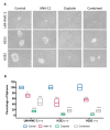
(A) Representative images of tumour spheroid under phase contrast microscopy. Note that cells under Cisplatin treatment have no ability to form well-developed spheres, however, in a combined group, this ability is recovered. (B) Quantitative analysis revealed that all treatments significantly reduced the number of spheres, however, it is possible to notice an increase in combined treatment compared to isolated Cisplatin (** p < 0.01; *** p < 0.001; **** p < 0.0001).

References

    1. Ferlay J., Ervik M., Lam F., Colombet M., Mery L., Piñeros M., Znaor A., Soerjomataram I., Bray F. Global Cancer Observatory: Cancer Tomorrow. International Agency for Research on Cancer; Lyon, France: 2018. [(accessed on 5 February 2020)]. Available online: https://gco.iarc.fr/tomorrow.
    1. El-Naggar A.K., Chan J.K.C., Grandis J.R., Takata T., Slootweg P.J. WHO Classification of Head and Neck Tumours. 4th ed. Volume 9 IARC; Lyon, France: 2017.
    1. Fonseca F.P., Carvalho Mde V., de Almeida O.P., Rangel A.L., Takizawa M.C., Bueno A.G., Vargas P.A. Clinicopathologic analysis of 493 cases of salivary gland tumours in a Southern Brazilian population. Oral Surg. Oral Med. Oral Pathol. Oral Radiol. 2012;114:230–239. doi: 10.1016/j.oooo.2012.04.008. - DOI - PubMed
    1. Jones A., Craig G., Speight P.M., Franklin C. The range and demographics of salivary gland tumours diagnosed in a UK population. Oral Oncol. 2008;44:407–417. doi: 10.1016/j.oraloncology.2007.05.010. - DOI - PubMed
    1. Huo Z., Wu H., Li J., Li S., Wu S., Liu Y., Luo Y., Cao J., Zeng X., Liang Z. Primary Pulmonary Mucoepidermoid Carcinoma: Histopathological and Moleculargenetic Studies of 26 Cases. PLoS ONE. 2015;10:e0143169. doi: 10.1371/journal.pone.0143169. - DOI - PMC - PubMed

LinkOut - more resources