Review
. 2020 Nov 24;9(12):2536.
doi: 10.3390/cells9122536.

Nutraceuticals and Exercise against Muscle Wasting during Cancer Cachexia

Affiliations
Review

Nutraceuticals and Exercise against Muscle Wasting during Cancer Cachexia

Giorgio Aquila et al. Cells. .

Abstract

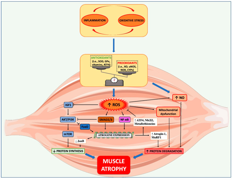
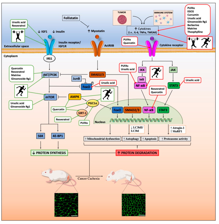
Cancer cachexia (CC) is a debilitating multifactorial syndrome, involving progressive deterioration and functional impairment of skeletal muscles. It affects about 80% of patients with advanced cancer and causes premature death. No causal therapy is available against CC. In the last few decades, our understanding of the mechanisms contributing to muscle wasting during cancer has markedly increased. Both inflammation and oxidative stress (OS) alter anabolic and catabolic signaling pathways mostly culminating with muscle depletion. Several preclinical studies have emphasized the beneficial roles of several classes of nutraceuticals and modes of physical exercise, but their efficacy in CC patients remains scant. The route of nutraceutical administration is critical to increase its bioavailability and achieve the desired anti-cachexia effects. Accumulating evidence suggests that a single therapy may not be enough, and a bimodal intervention (nutraceuticals plus exercise) may be a more effective treatment for CC. This review focuses on the current state of the field on the role of inflammation and OS in the pathogenesis of muscle atrophy during CC, and how nutraceuticals and physical activity may act synergistically to limit muscle wasting and dysfunction.

Keywords: bimodal approach; cancer cachexia; exercise; lifestyle interventions; muscle atrophy; muscle wasting; myokine; nutraceutical; nutrition.


Conflict of interest statement

The authors declare no conflict of interest. The funders had no role in the design of the study; in the collection, analyses, or interpretation of data; in the writing of the manuscript, or in the decision to publish the results.

Figures

Figure 1
Figure 1
Role of oxidative stress in muscle wasting during cancer cachexia. The crosstalk between increased inflammation and oxidative stress, occurring during cancer cachexia (CC), can drive in muscles an imbalance between antioxidant and pro-oxidant systems, favoring the generation of different sources of reactive oxygen species (ROS). During CC, ROS can reduce protein synthesis, by inhibiting IGF1/PI3K/mTOR pathway, and increase protein degradation, by stimulating pro-atrophic pathways such as FoxO3, SMAD2/3, and NF-κB. The latter promotes the ubiquitin proteasome system activation (atrogin-1 and MuRF1) that, along with ROS-mediated nitric oxide (NO) accumulation and mitochondrial dysfunction, facilitates the hypercatabolism typical of CC. Despite the concomitant activation of detoxifying systems (ATF4, Nfe2l2, metallothioneins), OS seems not to be resolved in muscles during CC.
Figure 2
Figure 2
Beneficial effects of nutraceuticals and physical activity against muscle atrophy during cancer cachexia. Graphical representation of the main findings of in vitro and in vivo studies, which have investigated the mechanisms underlying the benefits of the nutraceuticals and physical exercise against muscle wasting during cancer cachexia. In brief, nutraceuticals and physical exercise can favor protein synthesis in muscle by inducing both anabolic (i.e., IGF-1/AKT/mTOR) and anti-catabolic pathways (i.e., AMP-activated protein kinase (AMPK), SIRT1, and PGC1α) (in green). On the other hand, they can limit protein degradation by inhibiting pro-catabolic pathways (i.e., FoxO3, SMAD2/3, and NF-κB) (in red), thus limiting the expression of genes involved in mitochondrial dysfunction, autophagy, apoptosis, proteasomal degradation, and inflammation. The running man and the weightlifter represent “endurance/aerobic” and “resistance/strength” trainings, respectively. A transversal section of a tibialis anterior stained for wheat germ agglutinin (WGA) and coming from a healthy and a colon adenocarcinoma 26 (C26)-bearing mouse are shown. Scale bar: 100 μm.

References

    1. Tisdale M.J. Mechanisms of cancer cachexia. Physiol. Rev. 2009;89:381–410. doi: 10.1152/physrev.00016.2008. - DOI - PubMed
    1. Fearon K., Strasser F., Anker S.D., Bosaeus I., Bruera E., Fainsinger R.L., Jatoi A., Loprinzi C., MacDonald N., Mantovani G., et al. Definition and classification of cancer cachexia: An international consensus. Lancet Oncol. 2011;12:489–495. doi: 10.1016/S1470-2045(10)70218-7. - DOI - PubMed
    1. Miyamoto Y., Hanna D.L., Zhang W., Baba H., Lenz H.-J. Molecular Pathways: Cachexia Signaling–A Targeted Approach to Cancer Treatment. Clin. Cancer Res. 2016;22:3999–4004. doi: 10.1158/1078-0432.CCR-16-0495. - DOI - PMC - PubMed
    1. Houten L., Reilley A.A. An investigation of the cause of death from cancer. J. Surg. Oncol. 1980;13:111–116. doi: 10.1002/jso.2930130205. - DOI - PubMed
    1. Roberts B.M., Ahn B., Smuder A.J., Al-Rajhi M., Gill L.C., Beharry A.W., Powers S.K., Fuller D.D., Ferreira L.F., Judge A.R. Diaphragm and ventilatory dysfunction during cancer cachexia. FASEB J. 2013;27:2600–2610. doi: 10.1096/fj.12-222844. - DOI - PMC - PubMed

Publication types