

Hawthorn Herbal Preparation from Crataegus oxyacantha Attenuates In Vivo Carbon Tetrachloride -Induced Hepatic Fibrosis via Modulating Oxidative Stress and Inflammation
- PMID: 33255507
- PMCID: PMC7760839
- DOI: 10.3390/antiox9121173
Hawthorn Herbal Preparation from Crataegus oxyacantha Attenuates In Vivo Carbon Tetrachloride -Induced Hepatic Fibrosis via Modulating Oxidative Stress and Inflammation
Abstract
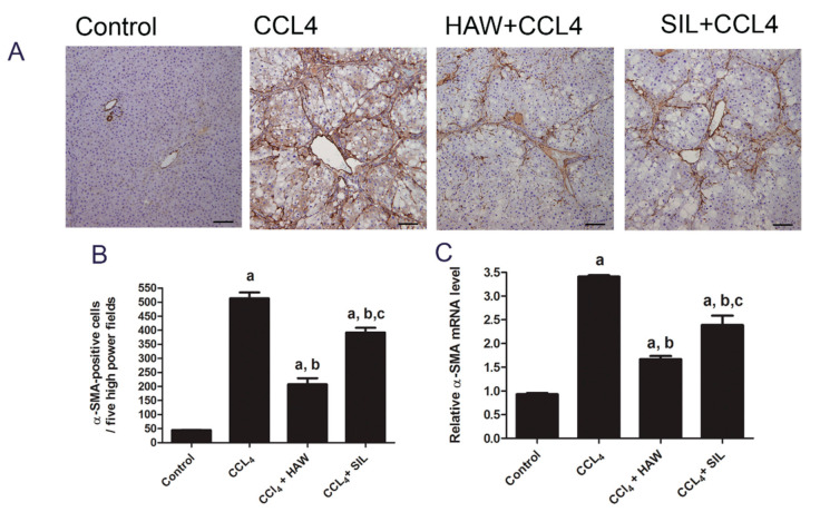
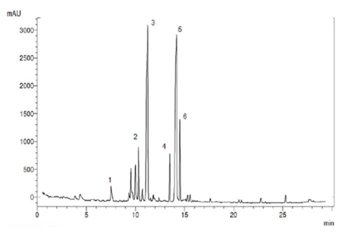
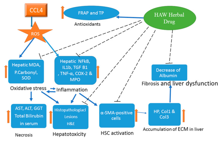
Hawthorn (HAW) is a herbal preparation extracted from Crataegus oxyacantha. HAW has cardioprotective, antioxidants, anti-inflammatory, and anti-hypotensive effects. HAW's effect on hepatic fibrosis remains, however, unknown. This study evaluated the impact of HAW on carbon tetrachloride (CCl4)-induced hepatic fibrosis in rats and elucidated its mechanisms. HAW reduced liver index and the serum liver enzyme markers and reduced liver damage, and fibrosis as confirmed by histopathological scoring of hematoxylin-eosin staining. Collagen deposition was reduced in HAW group compared to CCl4 group as confirmed by Masson staining, hydroxyproline content, and both mRNA and protein levels of alpha-smooth muscle actin, collagen 1 and 3. HAW also down regulated the gene expressions of inflammatory markers including interleukin-IL-1β, tumor necrosis factor-α, transforming growth factor-β 1, nuclear factor kappa-B, and cyclooxygenase-2 and decreased the myeloperoxidase activity. The effects of HAW was also associated with decreased levels of hepatic oxidative stress markers (malondialdehyde and P.Carbonyl) and with increased activity of superoxide dismutase. Those effects are possibly mediated by blocking the pro-oxidant machinery and down regulating the inflammatory and profibrotic responses. Finally, chlorogenic acid, epicatechin, rutin, vitexin quercetin, and iso quercetin were identified as the major species of polyphenols of the HAW herbal preparation used here. Therefore, HAW's potent protecting effects against liver fibrosis predicts a significant beneficial application.
Keywords: antioxidants; carbon tetrachloride; hawthorn; liver fibrosis.
Conflict of interest statement
The authors declare no conflict of interest.
Figures


Similar articles
-
Stachydrine ameliorates carbon tetrachloride-induced hepatic fibrosis by inhibiting inflammation, oxidative stress and regulating MMPs/TIMPs system in rats.Biomed Pharmacother. 2018 Jan;97:1586-1594. doi: 10.1016/j.biopha.2017.11.117. Epub 2017 Nov 27. Biomed Pharmacother. 2018. PMID: 29378386
-
Antifibrotic effect of meloxicam in rat liver: role of nuclear factor kappa B, proinflammatory cytokines, and oxidative stress.Naunyn Schmiedebergs Arch Pharmacol. 2016 Sep;389(9):971-83. doi: 10.1007/s00210-016-1263-1. Epub 2016 May 31. Naunyn Schmiedebergs Arch Pharmacol. 2016. PMID: 27245167
-
Hepatoprotective Effects of Grape Seed Procyanidin B2 in Rats With Carbon Tetrachloride-induced Hepatic Fibrosis.Altern Ther Health Med. 2015;21 Suppl 2:12-21. Altern Ther Health Med. 2015. PMID: 26308756
-
Dimethylfumarate ameliorates hepatic injury and fibrosis induced by carbon tetrachloride.Chem Biol Interact. 2019 Apr 1;302:53-60. doi: 10.1016/j.cbi.2019.01.029. Epub 2019 Jan 28. Chem Biol Interact. 2019. PMID: 30703375
-
Effect of crataegus usage in cardiovascular disease prevention: an evidence-based approach.Evid Based Complement Alternat Med. 2013;2013:149363. doi: 10.1155/2013/149363. Epub 2013 Dec 29. Evid Based Complement Alternat Med. 2013. PMID: 24459528 Free PMC article. Review.
Cited by
-
Advances in neovascularization after diabetic ischemia.World J Diabetes. 2022 Nov 15;13(11):926-939. doi: 10.4239/wjd.v13.i11.926. World J Diabetes. 2022. PMID: 36437864 Free PMC article. Review.
-
Tamarix articulata Induced Prevention of Hepatotoxicity Effects of In Vivo Carbon Tetrachloride by Modulating Pro-Inflammatory Serum and Antioxidant Enzymes to Reverse the Liver Fibrosis.Antioxidants (Basel). 2022 Sep 15;11(9):1824. doi: 10.3390/antiox11091824. Antioxidants (Basel). 2022. PMID: 36139897 Free PMC article.
-
Botanical, Phytochemical, Anti-Microbial and Pharmaceutical Characteristics of Hawthorn (Crataegusmonogyna Jacq.), Rosaceae.Molecules. 2021 Nov 30;26(23):7266. doi: 10.3390/molecules26237266. Molecules. 2021. PMID: 34885847 Free PMC article. Review.
-
The Role of Pyrazolo[3,4-d]pyrimidine-Based Kinase Inhibitors in The Attenuation of CCl4-Induced Liver Fibrosis in Rats.Antioxidants (Basel). 2023 Mar 3;12(3):637. doi: 10.3390/antiox12030637. Antioxidants (Basel). 2023. PMID: 36978885 Free PMC article.
-
Traditional Chinese medicine: An important source for discovering candidate agents against hepatic fibrosis.Front Pharmacol. 2022 Aug 23;13:962525. doi: 10.3389/fphar.2022.962525. eCollection 2022. Front Pharmacol. 2022. PMID: 36081936 Free PMC article. Review.
References
LinkOut - more resources
Full Text Sources
Research Materials
