Review
. 2020 Nov 24;21(23):8922.
doi: 10.3390/ijms21238922.

WWOX Loss of Function in Neurodevelopmental and Neurodegenerative Disorders

Affiliations
Review

WWOX Loss of Function in Neurodevelopmental and Neurodegenerative Disorders

C Marcelo Aldaz et al. Int J Mol Sci. .

Abstract

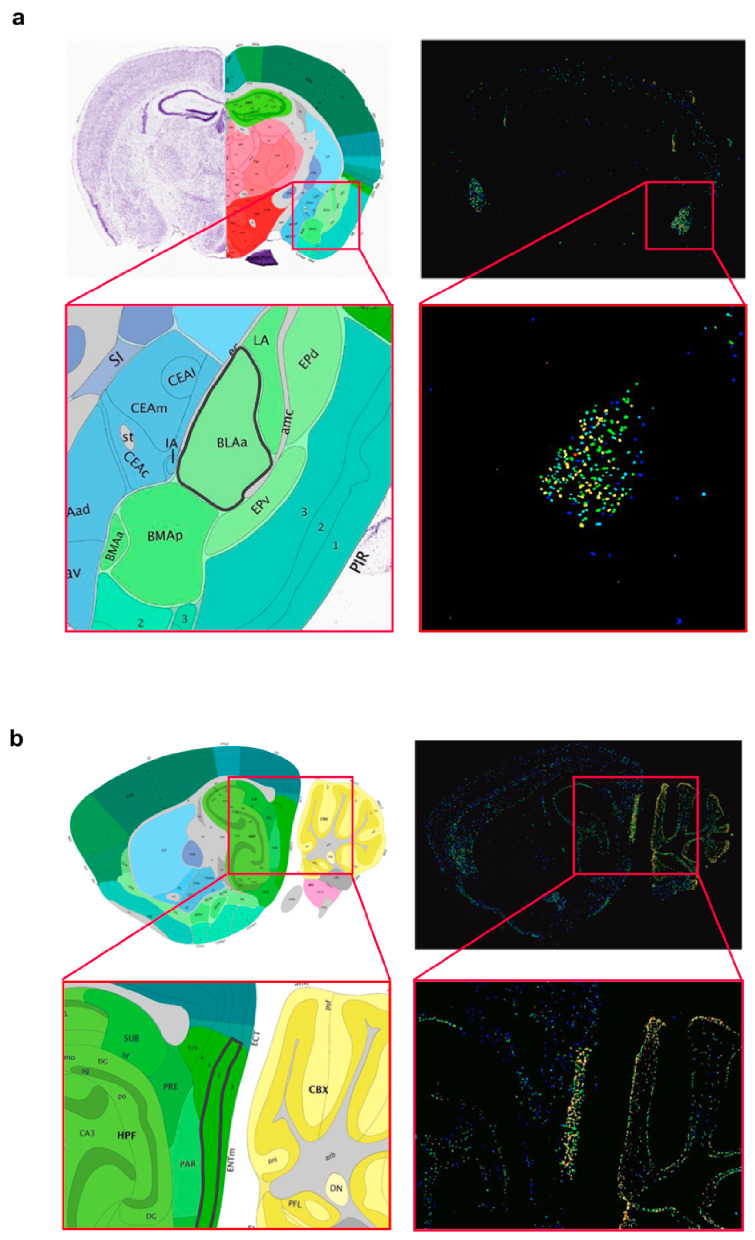
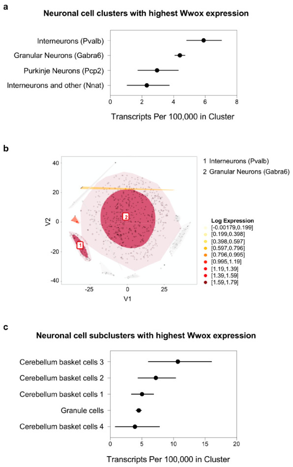
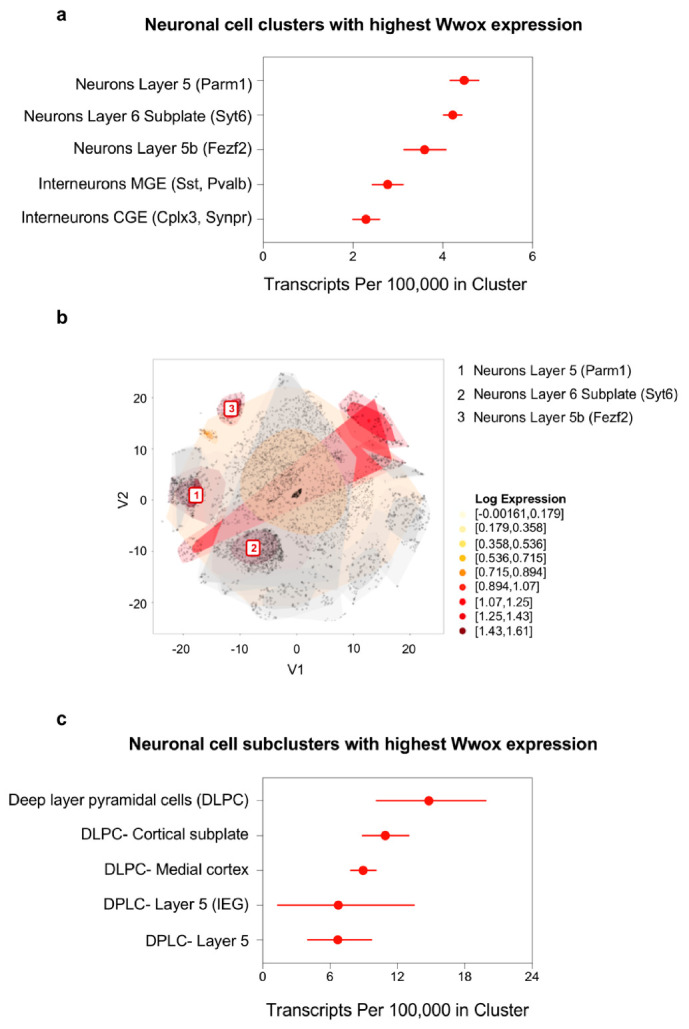
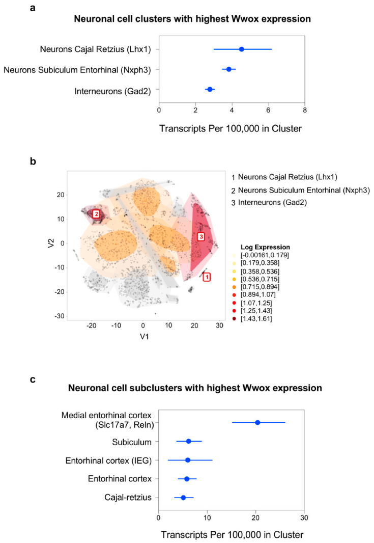
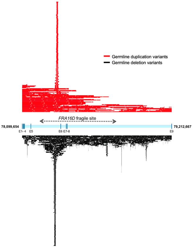
The WWOX gene was initially discovered as a putative tumor suppressor. More recently, its association with multiple central nervous system (CNS) pathologies has been recognized. WWOX biallelic germline pathogenic variants have been implicated in spinocerebellar ataxia type 12 (SCAR12; MIM:614322) and in early infantile epileptic encephalopathy (EIEE28; MIM:616211). WWOX germline copy number variants have also been associated with autism spectrum disorder (ASD). All identified germline genomic variants lead to partial or complete loss of WWOX function. Importantly, large-scale genome-wide association studies have also identified WWOX as a risk gene for common neurodegenerative conditions such as Alzheimer's disease (AD) and multiple sclerosis (MS). Thus, the spectrum of CNS disorders associated with WWOX is broad and heterogeneous, and there is little understanding of potential mechanisms at play. Exploration of gene expression databases indicates that WWOX expression is comparatively higher in the human cerebellar cortex than in other CNS structures. However, RNA in-situ hybridization data from the Allen Mouse Brain Atlas show that specific regions of the basolateral amygdala (BLA), the medial entorhinal cortex (EC), and deep layers of the isocortex can be singled out as brain regions with specific higher levels of Wwox expression. These observations are in close agreement with single-cell RNA-seq data which indicate that neurons from the medial entorhinal cortex, Layer 5 from the frontal cortex as well as GABAergic basket cells and granule cells from cerebellar cortex are the specific neuronal subtypes that display the highest Wwox expression levels. Importantly, the brain regions and cell types in which WWOX is most abundantly expressed, such as the EC and BLA, are intimately linked to pathologies and syndromic conditions in turn associated with this gene, such as epilepsy, intellectual disability, ASD, and AD. Higher Wwox expression in interneurons and granule cells from cerebellum points to a direct link to the described cerebellar ataxia in cases of WWOX loss of function. We now know that total or partial impairment of WWOX function results in a wide and heterogeneous variety of neurodegenerative conditions for which the specific molecular mechanisms remain to be deciphered. Nevertheless, these observations indicate an important functional role for WWOX in normal development and function of the CNS. Evidence also indicates that disruption of WWOX expression at the gene or protein level in CNS has significant deleterious consequences.

Keywords: ADHD; Alzheimer’s disease; WOREE; WWOX; autism; epileptic encephalopathy; intellectual disability; multiple sclerosis; neurodegeneration; spinocerebellar ataxia.


Conflict of interest statement

The authors declare no conflict of interest.

Figures

Figure 1
Figure 1
Temporospatial WWOX expression in human CNS tissues. (a) Trajectory plot showing the expression of WWOX during fetal development (period 1–7), infancy (period 8–9), childhood (period 10–11), adolescence (period 12), and adulthood (period 13–15), in individual CNS areas: neocortex (NCX), hippocampus (HIP), amygdala region (AMY), striatum (STR), mediodorsal nucleus of the thalamus (MD), and cerebellar cortex (CBC). Reprinted and adapted from Human Brain Transcriptome dataset (https://hbatlas.org/) [17]. (b) Violin plots showing the expression of WWOX in different brain regions as per the Genotype-Tissue Expression dataset (GTEx, Analysis Release V8, dbGaP Accession phs000424.v8.p2). Horizontal line in black box indicates the median value of WWOX expression in respective tissues; the number of samples for each tissue is indicated in the X-axis legend. TPM: Transcripts per Million. Reprinted and adapted from the RNA-seq GTEx database (https://gtexportal.org) [18].
Figure 2
Figure 2
Localized Wwox expression in basolateral amygdala and medial entorhinal cortex of mouse brain. (a) Coronal brain section showing Wwox mRNA in-situ hybridization signals clearly delineating the anterior (BLAa) region of the basolateral amigdalar nucleus in the cortical subplate. The inset shows the zoomed image of the BLAa region, highlighted with a black border in the annotated panel (left). (b) Sagittal section showing in situ hybridization signals, specifically lighting-up layer 2 of the medial entorhinal cortex (ENTm2, medial part, dorsal zone, layer 2). The inset shows the zoomed image of the ENTm2 region, highlighted with a black border in the annotated panel (left). It can also be observed that Wwox expression clearly delineates specific layers of the cerebellar cortex. Brain tissue sections from 56-day old, C57Bl/6J male mouse. All images were obtained from the Allen Mouse Brain Atlas—Allen Institute (https://mouse.brain-map.org/) [19].
Figure 3
Figure 3
Wwox expression in mouse brain cell types. Bar graphs representing Wwox cell-type-specific expression levels, obtained from a mouse brain RNA-seq database (www.BrainRNAseq.org) [22,23] in (a) neurons, microglia, astrocytes, and (b) oligodendrocytes. Within the oligodendrocyte population, the highest Wwox expression levels are seen in progenitor oligodendrocytes, followed by newly formed and myelinated oligodendrocytes. (c) Uniform Wwox expression is observed at P7, P21, and P60 in microglial cells. (d) Wwox expression is upregulated upon LPS treatment. Y-axis represents Wwox expression as fragments per kilobase of transcript per million mapped reads (FPKM), error bars represent ± SD.
Figure 4
Figure 4
Wwox expression at single-cell level in mouse cerebellum neurons. (a) Graph representing neuronal cell clusters. Pvalb+ interneurons and Gabra6+ granule cells express the most Wwox transcripts in mouse cerebellum. Markers specific to each cell cluster are shown in parenthesis. (b) t-SNE plot highlights the top two clusters described in (a) as the darkest colored regions among cerebellum cells. (c) Graph representing specific neuronal subclusters expressing the most Wwox transcripts. GABAergic basket cells (i.e., interneurons) and granular neurons are the top cell types. The confidence intervals in graphs (a,c) reflect statistical sampling noise calculated from the binomial distribution and reflecting the total number of unique molecular identifier (UMIs) ascertained by cluster rather than cell-to-cell heterogeneity within a cluster. Data was obtained from the mouse brain single-cell RNA sequencing (scRNA-seq) DropViz database (http://dropviz.org/) [28].
Figure 5
Figure 5
Wwox expression at single-cell level in mouse frontal cortex neurons. (a) Graph representing the neuronal cell clusters expressing the highest number of Wwox transcripts in mouse frontal cortex. Markers specific to each cell cluster are shown in parenthesis. Markers Parm1, Syt6 and Fezf2 identify the top three clusters. (b) t-SNE plot shows these three clusters as the darkest colored regions. (c) Graph representing the frontal cortex subclusters expressing the most Wwox transcripts, specifically deep-layer pyramidal neurons. The confidence intervals in graphs (a,c) reflect statistical sampling noise calculated from the binomial distribution and show the total number of UMIs ascertained by cluster. Data was obtained from the mouse brain scRNA-seq DropViz database (http://dropviz.org/) [28].
Figure 6
Figure 6
Wwox expression at single-cell level in mouse hippocampal neurons. (a) Graph representing the top three neuronal cell clusters expressing the highest number of Wwox transcripts identified by markers Lhx1, Nxph3 and Gad2 in mouse hippocampus. (b) These three clusters are shown in the t-SNE plot as the darkest colored regions. (c) Graph representing the hippocampal subclusters shows that neurons from the medial entorhinal cortex (markers Slc17a7 and Reln) express the most Wwox transcripts. The confidence intervals in graphs (a,c) reflect statistical sampling noise calculated from the binomial distribution and show the total number of UMIs ascertained by cluster. Data was obtained from the mouse brain scRNA-seq DropViz database (http://dropviz.org/) [28].
Figure 7
Figure 7
WWOX germline pathogenic variants in SCAR12 and WWOX-related epileptic encephalopathy (WOREE). (a) Missense, nonsense, and splice-site/intronic variants affecting WWOX protein in SCAR12 (red colored circles) and WOREE cases (gray colored circles for amino acid alterations and blue colored circles for splice-site/intronic variants). Size of each circle corresponds to the frequency of occurrence of the specific variant in single or multiple families, as noted. Mutation hotspots with more than one mutation at the same amino acid site or variants identified in more than one family are noted in red text. (b) Mapping of germline CNVs, duplications shown in red and deletions in black, of the WWOX locus in WOREE cases. Numbers next to dotted lines with arrowhead indicate coordinates of chromosomal breakpoints beyond the WWOX locus (human genome assembly GRCh38/hg38).
Figure 8
Figure 8
WWOX germline copy number variants (CNVs) in autism spectrum disorder (ASD). Distribution of WWOX locus CNVs associated with ASD and ID cases obtained from the AutDB database (http://www.mindspec.org/autdb.html). CNVs are mostly intragenic; however, additional larger variants with only one of the breakpoints within the genomic region spanned by this gene are also observed. Numbers next to dotted lines with arrowhead indicate coordinates of chromosomal breakpoints beyond the WWOX locus (human genome assembly GRCh38/hg38). Numbers 1–49 shown next to CNV bars correspond to case numbers described in Table S2.
Figure 9
Figure 9
WWOX is a hotspot for germline CNV polymorphisms in healthy humans. Mapping of non-redundant WWOX intragenic germline duplications (red) and deletions (black) variants in normal human population. Larger CNVs spanning beyond WWOX with one of the breakpoints within the gene are also shown. As can be observed there is a significant accumulation of germline CNVs clustering in a specific hotspot within intron 5. This hotspot overlaps the 5′ prime edge of the core of FRA16D (dotted line).

Similar articles

Cited by

References

    1. Bednarek A.K., Laflin K.J., Daniel R.L., Liao Q., Hawkins K.A., Aldaz C.M. WWOX, a novel WW domain-containing protein mapping to human chromosome 16q23.3-24.1, a region frequently affected in breast cancer. Cancer Res. 2000;60:2140–2145. - PubMed
    1. Bednarek A.K., Keck-Waggoner C.L., Daniel R.L., Laflin K.J., Bergsagel P.L., Kiguchi K., Brenner A.J., Aldaz C.M. WWOX, the FRA16D gene, behaves as a suppressor of tumor growth. Cancer Res. 2001;61:8068–8073. - PubMed
    1. Aldaz C.M., Ferguson B.W., Abba M.C. WWOX at the crossroads of cancer, metabolic syndrome related traits and CNS pathologies. Biochim. Biophys. Acta. 2014;1846:188–200. doi: 10.1016/j.bbcan.2014.06.001. - DOI - PMC - PubMed
    1. Schrock M.S., Batar B., Lee J., Druck T., Ferguson B., Cho J.H., Akakpo K., Hagrass H., Heerema N.A., Xia F., et al. Wwox-Brca1 interaction: Role in DNA repair pathway choice. Oncogene. 2017;36:2215–2227. doi: 10.1038/onc.2016.389. - DOI - PMC - PubMed
    1. Mallaret M., Synofzik M., Lee J., Sagum C.A., Mahajnah M., Sharkia R., Drouot N., Renaud M., Klein F.A., Anheim M., et al. The tumour suppressor gene WWOX is mutated in autosomal recessive cerebellar ataxia with epilepsy and mental retardation. Brain. 2014;137:411–419. doi: 10.1093/brain/awt338. - DOI - PMC - PubMed

MeSH terms

Substances