. 2020 Nov 25;12(12):1138.
doi: 10.3390/pharmaceutics12121138.

Optimization of Liposomes for Antigen Targeting to Splenic CD169+ Macrophages

Affiliations

Optimization of Liposomes for Antigen Targeting to Splenic CD169+ Macrophages

Maarten K Nijen Twilhaar et al. Pharmaceutics. .

Abstract

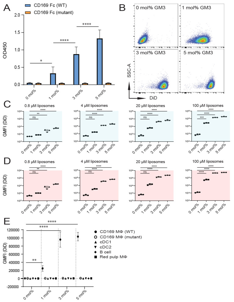
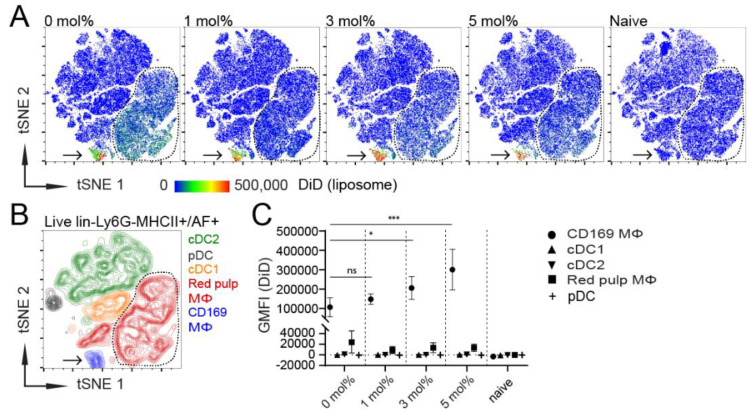
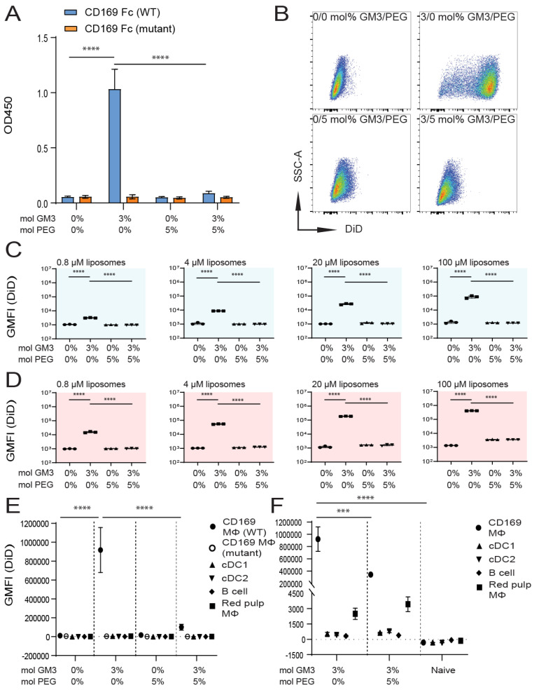
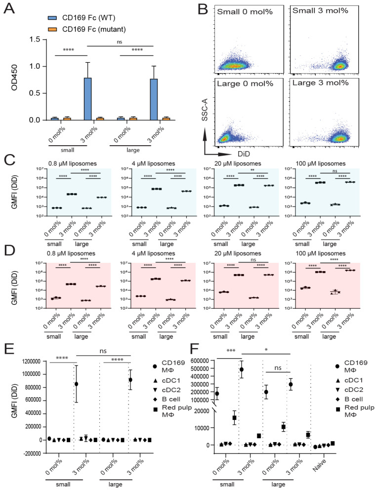
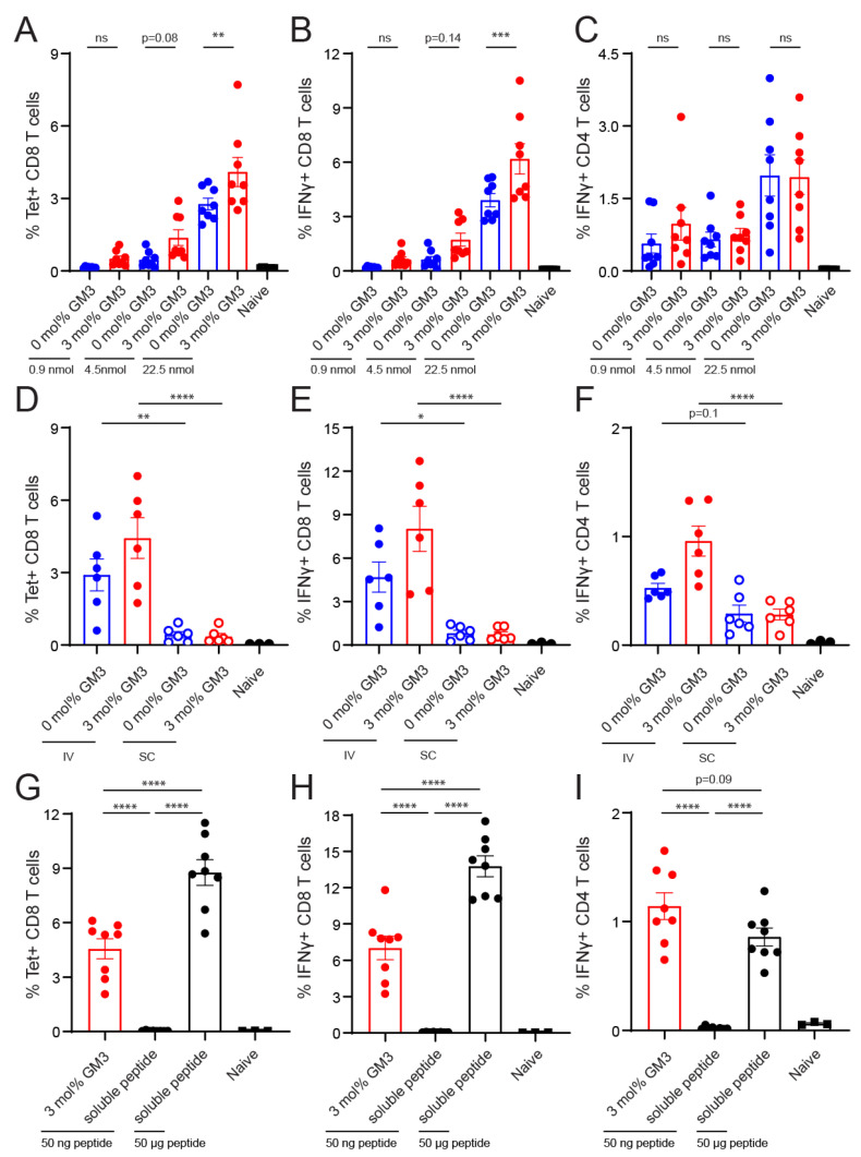
Despite promising progress in cancer vaccination, therapeutic effectiveness is often insufficient. Cancer vaccine effectiveness could be enhanced by targeting vaccine antigens to antigen-presenting cells, thereby increasing T-cell activation. CD169-expressing splenic macrophages efficiently capture particulate antigens from the blood and transfer these antigens to dendritic cells for the activation of CD8+ T cells. In this study, we incorporated a physiological ligand for CD169, the ganglioside GM3, into liposomes to enhance liposome uptake by CD169+ macrophages. We assessed how variation in the amount of GM3, surface-attached PEG and liposomal size affected the binding to, and uptake by, CD169+ macrophages in vitro and in vivo. As a proof of concept, we prepared GM3-targeted liposomes containing a long synthetic ovalbumin peptide and tested the capacity of these liposomes to induce CD8+ and CD4+ T-cell responses compared to control liposomes or soluble peptide. The data indicate that the delivery of liposomes to splenic CD169+ macrophages can be optimized by the selection of liposomal constituents and liposomal size. Moreover, optimized GM3-mediated liposomal targeting to CD169+ macrophages induces potent immune responses and therefore presents as an interesting delivery strategy for cancer vaccination.

Keywords: CD169; GM3; Siglec-1; T cells; cancer vaccination; liposome; macrophage; sialoadhesin; targeting.


Conflict of interest statement

The authors declare no conflict of interest.

Figures

Figure 1
Figure 1
Inclusion of GM3 in liposomes results in specific binding to CD169-expressing cells in vitro. (A) Liposomes were coated overnight on an ELISA plate, and binding to wild-type (WT) or mutant mouse CD169 Fc was quantified. Indicated is the average OD450 of 3 independent experiments. (BD) 1′-dioctadecyl-3,3,3′,3′-tetramethyl indodicarbocyanine (DiD)-containing liposomes were incubated with TSn at 4 or 37 °C for 45 min. Representative dot plots following incubation on 37 °C (20 µM liposomes) (B). Indicated is the average geometric mean fluorescence intensity GMFI ± SD of a technical triplicate after incubation at 4 °C (C) and at 37 °C (D) (pattern representative of 4 independent experiments). (E) DiD-containing liposomes were incubated for 45 min at 37 °C with freshly isolated splenocytes from C57BL/6 WT or W2QR97A-mutant CD169 mice. Subsequently, DiD fluorescence was quantified for distinct cell populations using flow cytometry. Indicated is the GMFI ± SD (n = 5) (representative of 2 independent experiments). * p < 0.05, ** p <0.01, *** p < 0.005 and **** p < 0.0001, ns: no significance.
Figure 2
Figure 2
CD169+ MΦ are the main antigen-presenting cells (APCs) that take up liposomes, and the inclusion of 3–5 mol% GM3 augments targeting efficiency. (A) DiD-containing liposomes (22.5 nmol of phospholipid), supplemented with adjuvant, were intravenously (IV) injected into mice. After 2 h, splenocytes were stained and major splenic APC populations were clustered using high-dimensional data reduction analysis (pre-gated on live, lineage, Ly6G and MHCII+/AF+ cells). Liposomal DiD fluorescence in APC populations is depicted for 0, 1, 3 or 5 mol% GM3-containing liposomes and non-injected naive mice. (B) CD169+ MΦ, red pulp MΦ, cDC1, cDC2 and pDC were manually gated and overlaid to identify the clusters that were obtained by high-dimensional data reduction. Arrow points to CD169+ MΦ, and red pulp MΦ are bordered by a line. (C) Quantification of DiD fluorescence within various splenic APC populations. Indicated is the average GMFI ± SD (n = 6 for CD169+ MΦ, red pulp MΦ, cDC1 and cDC2 and n = 3 for pDC). * p < 0.05, *** p < 0.001, ns: no significance.
Figure 3
Figure 3
Liposomal incorporation of PEG abolishes the interaction between GM3 and CD169. (A) Liposomes were coated overnight on an ELISA plate, and binding to WT or mutant CD169 Fc was quantified. Indicated is the average OD450 of 3 independent experiments. (BD) DiD-containing liposomes were incubated with TSn at 4 or 37 °C for 45 min. Representative dot plots following incubation at 37 °C (20 µM liposomes) (B). Indicated is the average GMFI ± SD of technical triplicates after incubation at 4 °C (C) and at 37 °C (D) (pattern representative of 4 independent experiments). (E) DiD-containing liposomes were incubated for 45 min at 37 °C with freshly isolated splenocytes from C57BL/6 WT or W2QR97A-mutant CD169 mice. Subsequently, DiD fluorescence was quantified for distinct cell populations using flow cytometry. Indicated is the GMFI ± SD (n = 5) (representative of 2 independent experiments). (F) DiD-containing liposomes (90 nmol of phospholipid), supplemented with adjuvant, were injected IV in mice, and after 2 h, splenic cell populations were analyzed for DiD fluorescence. Indicated is the average GMFI ± SD (n = 4). *** p < 0.005 and **** p < 0.0001, ns: no significance.
Figure 4
Figure 4
Size of GM3-containing liposomes influences binding to CD169-expressing cells in vivo, but not in vitro. (A) Liposomes were coated overnight on an ELISA plate, and binding to WT or mutant CD169 Fc was quantified. Indicated is the average OD450 of 2 independent technical triplicates. (BD) DiD-containing liposomes were incubated with TSn at 4 or 37 °C for 45 min. Representative dot plots following incubation at 37 °C (20 µM liposomes) (B). Indicated is the average GMFI ± SD of a technical triplicate after incubation at 4 °C (C) and at 37 °C (D) (representative of 4 independent experiments). (E) DiD-containing liposomes were incubated for 45 min at 37 °C with freshly isolated splenocytes from C57BL/6 WT mice. Subsequently, DiD fluorescence was quantified for distinct cell populations using flow cytometry. Indicated is the GMFI ± SD (n = 4) (representative of 2 independent experiments). (F) DiD-containing liposomes (22.5 nmol of phospholipid) were injected IV in mice, and after 2 h, splenic cell populations were analyzed for DiD fluorescence. Indicated is the average GMFI ± SD (n = 4) (F). * p < 0.05, ** p < 0.01, *** p < 0.005 and **** p < 0.0001, ns: no significance.
Figure 5
Figure 5
Intravenous administration of GM3-containing liposomes augments vaccine effectiveness. (AC) Liposomes were IV administered to mice (22.5, 4.5 and 0.9 nmol of phospholipid for the high, middle and low doses, respectively), supplemented with adjuvant. (DF) Liposomes were IV or subcutaneously (SC) administered to mice (22.5 nmol of phospholipid), supplemented with adjuvant. (GI) GM3-containing liposomes with peptide (22.5 nmol of phospholipid, which equals 50 ng of peptide) or a low or high dose of free peptide (50 ng and 50 μg, respectively) were IV administered to mice, supplemented with adjuvant. (A,D,G) On Day 7, H-2Kb/SIINFEKL tetramer binding T cells were identified. (B,C,E,F,H,I) Splenocytes were restimulated with peptide for 5 h or 25 h, for CD8+ or CD4+ T-cell responses, respectively. Subsequently, IFNγ-producing CD8+ (B,E,H) or CD4+ (C,F,I) T cells were detected with intracellular flow cytometry staining. Indicated is the GMFI ± SEM (n = 8) (pooled from 2 independent experiments) for A–C, (n = 6) for the experimental and (n = 3) for the naïve group for D–F and (n = 8) for the experimental and (n = 3) for the naïve group for G–I. * p < 0.05, ** p < 0.01, *** p < 0.005 and **** p < 0.0001, ns: no significance.

Similar articles

Cited by

References

    1. Alexandrov L.B., Nik-Zainal S., Wedge D.C., Aparicio S.A.J.R., Behjati S., Biankin A.V., Bignell G.R., Bolli N., Borg A., Børresen-Dale A.-L., et al. Signatures of mutational processes in human cancer. Nature. 2013;500:415–421. doi: 10.1038/nature12477. - DOI - PMC - PubMed
    1. Schumacher T.N., Scheper W., Kvistborg P. Cancer Neoantigens. Annu. Rev. Immunol. 2019;37:173–200. doi: 10.1146/annurev-immunol-042617-053402. - DOI - PubMed
    1. Melief C.J., van Hall T., Arens R., Ossendorp F., van der Burg S.H. Therapeutic cancer vaccines. J. Clin. Investig. 2015;125:3401–3412. doi: 10.1172/JCI80009. - DOI - PMC - PubMed
    1. Haen S.P., Löffler M.W., Rammensee H.G., Brossart P. Towards new horizons: Characterization, classification and implications of the tumour antigenic repertoire. Nat. Rev. Clin. Oncol. 2020;17:595–610. doi: 10.1038/s41571-020-0387-x. - DOI - PMC - PubMed
    1. Pardoll D.M. The blockade of immune checkpoints in cancer immunotherapy. Nat. Rev. Cancer. 2012;12:252–264. doi: 10.1038/nrc3239. - DOI - PMC - PubMed