Review
. 2020 Nov 25;8(12):1856.
doi: 10.3390/microorganisms8121856.

Epidemiology and Sequence-Based Evolutionary Analysis of Circulating Non-Polio Enteroviruses

Affiliations
Review

Epidemiology and Sequence-Based Evolutionary Analysis of Circulating Non-Polio Enteroviruses

David M Brown et al. Microorganisms. .

Abstract

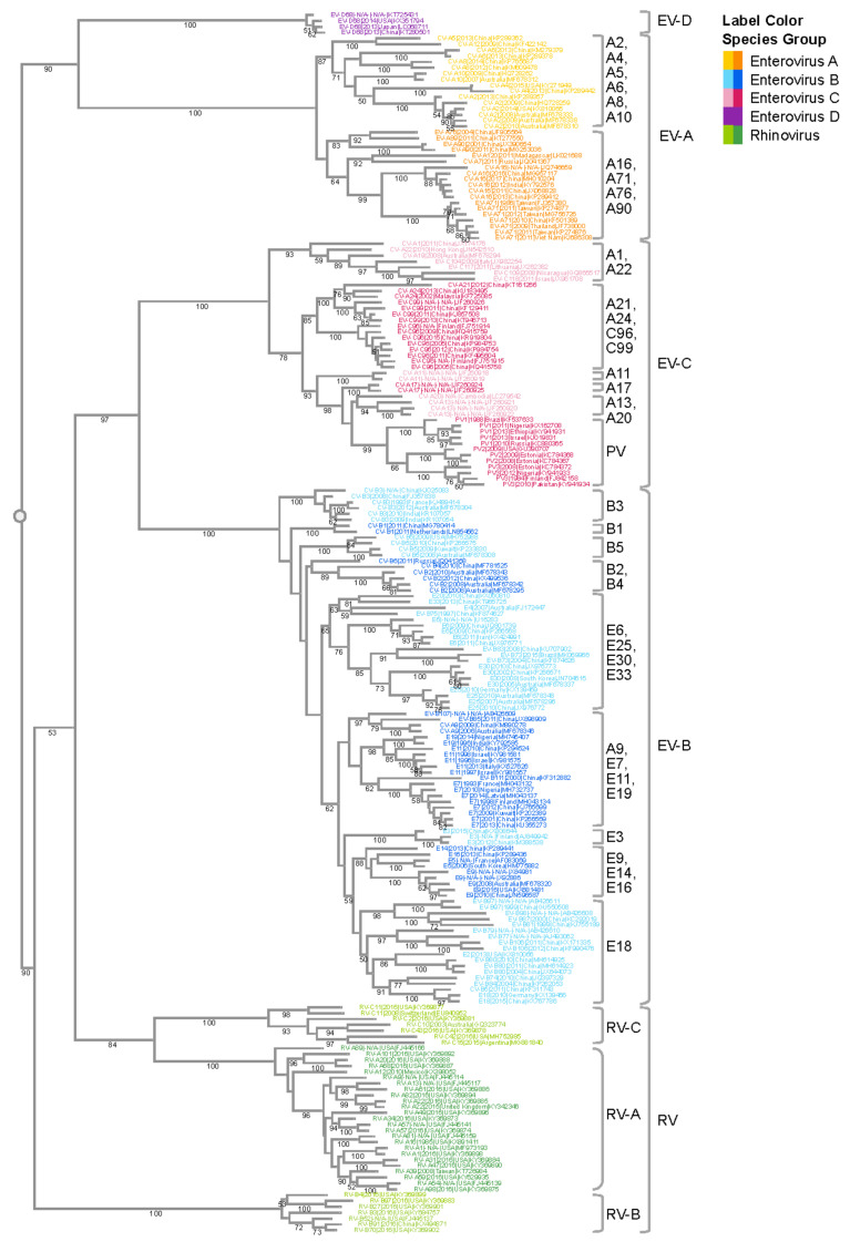
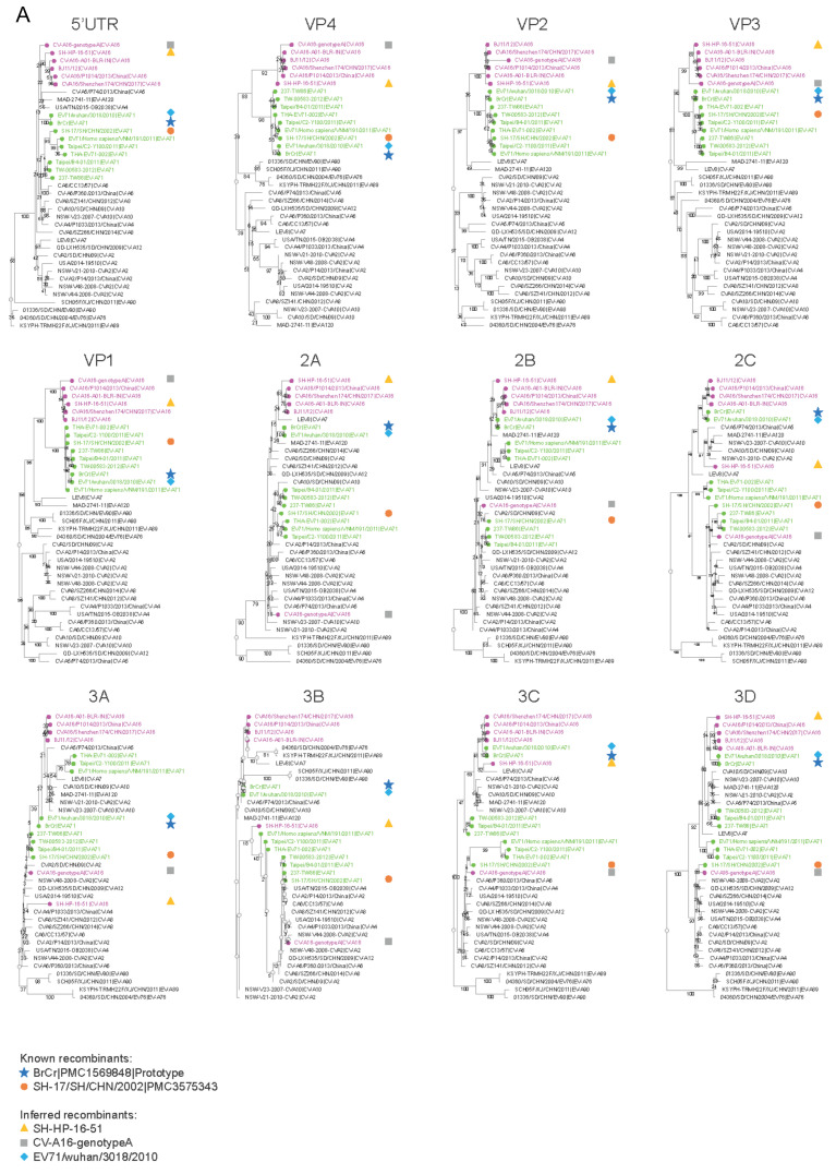
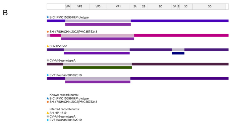
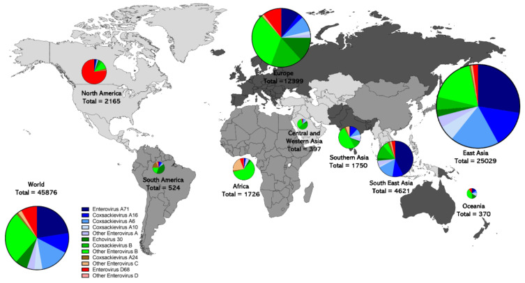
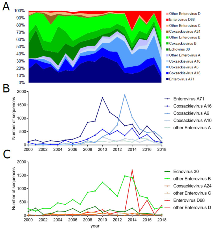
Enteroviruses (EVs) are positive-sense RNA viruses, with over 50,000 nucleotide sequences publicly available. While most human infections are typically associated with mild respiratory symptoms, several different EV types have also been associated with severe human disease, especially acute flaccid paralysis (AFP), particularly with endemic members of the EV-B species and two pandemic types-EV-A71 and EV-D68-that appear to be responsible for recent widespread outbreaks. Here we review the recent literature on the prevalence, characteristics, and circulation dynamics of different enterovirus types and combine this with an analysis of the sequence coverage of different EV types in public databases (e.g., the Virus Pathogen Resource). This evaluation reveals temporal and geographic differences in EV circulation and sequence distribution, highlighting recent EV outbreaks and revealing gaps in sequence coverage. Phylogenetic analysis of the EV genus shows the relatedness of different EV types. Recombination analysis of the EV-A species provides evidence for recombination as a mechanism of genomic diversification. The absence of broadly protective vaccines and effective antivirals makes human enteroviruses important pathogens of public health concern.

Keywords: Echovirus 30 (E30); Virus Pathogen Resource (ViPR); acute flaccid myelitis (AFM); acute flaccid paralysis (AFP); and mouth disease (HFMD); coxsackievirus A16 (CV-A16); coxsackievirus A24 (CV-A24); coxsackievirus A6 (CV-A6); enterovirus A71 (EV-A71); enterovirus D68 (EV-D68); foot; hand.


Conflict of interest statement

The authors declare no conflict of interest. The sponsors had no role in the design, execution, interpretation, or writing of the study/review.

Figures

Figure 1
Figure 1
Phylogenetic analysis of the enterovirus genus. VP1 gene sequences from complete human enterovirus genomes were retrieved from the ViPR database (https://www.viprbrc.org/). Sequences representative of the genomic diversity were selected using CD-HIT [128], with a threshold of 0.90, and subsequently aligned using MAFFT E-INS-i. The phylogenetic tree was generated using RaXML with 100 bootstrap replicates, and then visualized in Archaeopteryx.js on ViPR with leaf nodes color coded by phylogenetic relatedness. Bootstrap support values of 50 or larger are shown in the tree. Major monophyletic groups are labeled with the major types in the group. Major types are defined as those with 50 or more genomes in the case of EV-A and EV-C, and 200 or more genomes in the case of EV-B.
Figure 2
Figure 2
Recombination analysis of EV-A isolates. Gene sequences for the EV-A isolates selected in Figure 1 along with published recombinant strains BrCr (U22521) [136] and SH-17/SH/CHN/2002 (JX678885) [137] were analyzed. (A) Phylogenetic trees of different genomic regions. Individual gene sequences were retrieved from the ViPR website (https://www.viprbrc.org/). Trees were generated using RaXML with 100 bootstrap replicates, and subsequently visualized in Archaeopteryx.js on ViPR. CV-A16 strains are labeled in magenta; EV-A71 strains are labeled in green. Recombinant controls (BrCr, SH-17/SH/CHN/2002) are highlighted by navy and orange shapes, respectively. Three novel recombinants (SH-HP-16-51, CV-A16-genotypeA, EV71/wuhan/3018/2010) were inferred based on changes in their phylogenetic neighborhoods between individual genes and are highlighted by yellow, gray, and light blue shapes. (B) Recombination analysis of the same dataset using RDP4 [138] detected recombination events in recombinant controls (BrCr, SH-17/SH/CHN/2002) and the three inferred recombinants (SH-HP-16-51, CV-A16-genotypeA, EV71/wuhan/3018/2010) in regions consistent with the phylogenetic analysis.
Figure 2
Figure 2
Recombination analysis of EV-A isolates. Gene sequences for the EV-A isolates selected in Figure 1 along with published recombinant strains BrCr (U22521) [136] and SH-17/SH/CHN/2002 (JX678885) [137] were analyzed. (A) Phylogenetic trees of different genomic regions. Individual gene sequences were retrieved from the ViPR website (https://www.viprbrc.org/). Trees were generated using RaXML with 100 bootstrap replicates, and subsequently visualized in Archaeopteryx.js on ViPR. CV-A16 strains are labeled in magenta; EV-A71 strains are labeled in green. Recombinant controls (BrCr, SH-17/SH/CHN/2002) are highlighted by navy and orange shapes, respectively. Three novel recombinants (SH-HP-16-51, CV-A16-genotypeA, EV71/wuhan/3018/2010) were inferred based on changes in their phylogenetic neighborhoods between individual genes and are highlighted by yellow, gray, and light blue shapes. (B) Recombination analysis of the same dataset using RDP4 [138] detected recombination events in recombinant controls (BrCr, SH-17/SH/CHN/2002) and the three inferred recombinants (SH-HP-16-51, CV-A16-genotypeA, EV71/wuhan/3018/2010) in regions consistent with the phylogenetic analysis.
Figure 3
Figure 3
Geographic distribution of non-polio enterovirus genomic sequence records. The origin (if available) of all non-polio enterovirus A (blue) B (green) C (brown) and D (red) sequences from samples collected from 2000 to 2018 are shown, highlighting the different distribution patterns of enterovirus types across nine different regions of the world. Data were obtained using the ViPR database, as described in Table 1 footnotes. Regional pie graph sizes are proportional to the square root of total sequences of each group.
Figure 4
Figure 4
Enterovirus sequence representation from 2000 to 2018 by type. The sample collection calendar year (if available) of all non-polio enteroviruses A (blue), B (green), C (brown), and D (red) sequences. The percentage of each type collected during each year is given in panel (A). The numbers of sequences collected from the most common types of EV-A and EV-B, -C, and -D are displayed in panels (B) and (C), respectively. Data were obtained using the ViPR database, as described in Table 1 footnotes.

Similar articles

Cited by

References

    1. Pallansch M.A., Roos R.P. Enteroviruses: Polioviruses, coxsackieviruses, echoviruses, and newer enteroviruses. In: Knipe D.M., Howley P.M., Griffin D.E., Lamb R.A., Martin M.A., Roizman B., Stephen E.S., editors. Fields Virology. 4th ed. Lippincott Williams & Wilkins; Philadelphia, PA, USA: 2001. pp. 723–775.
    1. Tangermann R.H., Aylward R., Birmingham M., Horner R., Olivé J.M., Nkowane B.M., Hull H.F., Burton A. Current status of the global eradication of poliomyelitis. World Health Stat. Q. Rapp. Trimest. Stat. Sanit. Mond. 1997;50:188–194. - PubMed
    1. Shaghaghi M., Soleyman-Jahi S., Abolhassani H., Yazdani R., Azizi G., Rezaei N., Barbouche M.-R., McKinlay M.A., Aghamohammadi A. New insights into physiopathology of immunodeficiency-associated vaccine-derived poliovirus infection; systematic review of over 5 decades of data. Vaccine. 2018;36:1711–1719. doi: 10.1016/j.vaccine.2018.02.059. - DOI - PubMed
    1. Guo J., Bolivar-Wagers S., Srinivas N., Holubar M., Maldonado Y. Immunodeficiency-related vaccine-derived poliovirus (iVDPV) cases: A systematic review and implications for polio eradication. Vaccine. 2015;33:1235–1242. doi: 10.1016/j.vaccine.2015.01.018. - DOI - PMC - PubMed
    1. Burns C.C., Diop O.M., Sutter R.W., Kew O.M. Vaccine-Derived Polioviruses. J. Infect. Dis. 2014;210:S283–S293. doi: 10.1093/infdis/jiu295. - DOI - PubMed