

Semen Modulates Inflammation and Angiogenesis in the Reproductive Tract of Female Rabbits
- PMID: 33255666
- PMCID: PMC7761520
- DOI: 10.3390/ani10122207
Semen Modulates Inflammation and Angiogenesis in the Reproductive Tract of Female Rabbits
Abstract
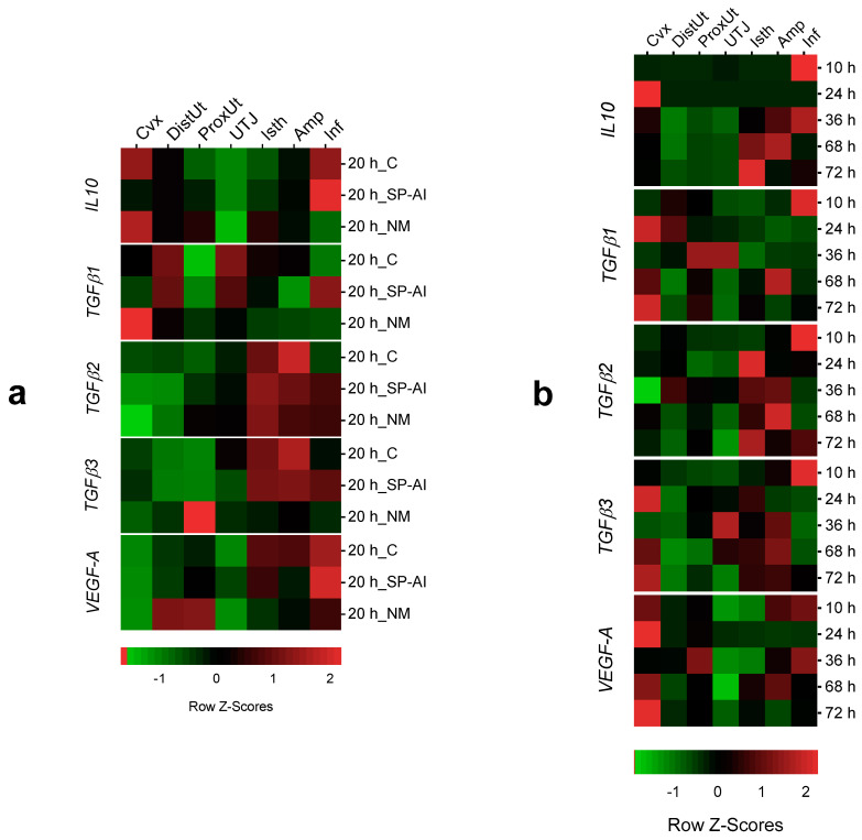
The maternal environment modulates immune responses to facilitate embryo development and ensure pregnancy. Unraveling this modulation could improve the livestock breeding systems. Here it is hypothesized that the exposure of the female rabbit reproductive tract to semen, as well as to early embryos, modulates inflammation and angiogenesis among different tissue segments. qPCR analysis of the gene expression changes of the anti-inflammatory interleukin-10 (IL10) and transforming growth factor beta family (TGFβ1-3) and the angiogenesis mediator vascular endothelial growth factor (VEGF-A) were examined in response to mating or insemination with sperm-free seminal plasma (SP). Reproductive tract segment (cervix to infundibulum) samples were obtained in Experiment 1, 20 h after gonadotropin-releasing hormone (GnRH) stimulation (control), natural mating (NM) or vaginal infusion with sperm-free SP (SP-AI). Additionally, segmented samples were also obtained at 10, 24, 36, 68 or 72 h after GnRH-stimulation and natural mating (Experiment 2). The results of gene expression, analyzed by quantitative PCR, showed that NM effects were mainly localized in the uterine tissues, depicting clear temporal variation, while SP-AI effects were restricted to the oviduct. Changes in anti-inflammatory and angiogenesis mediators indicate an early response in the uterus and a late modulation in the oviduct either induced by semen or preimplantation embryos. This knowledge could be used in the implementation of physiological strategies in breeding systems to face the new challenges on rabbit productivity and sustainability.
Keywords: angiogenesis; endometrium; gene expression; inflammation; oviduct; rabbit; seminal plasma; spermatozoa.
Conflict of interest statement
The authors declare no conflict of interest. The funders had no role in the design of the study; in the collection, analyses or interpretation of data; in the writing of the manuscript or in the decision to publish the results.
Figures


Similar articles
-
Seminal Plasma Triggers the Differential Expression of the Glucocorticoid Receptor (NR3C1/GR) in the Rabbit Reproductive Tract.Animals (Basel). 2020 Nov 19;10(11):2158. doi: 10.3390/ani10112158. Animals (Basel). 2020. PMID: 33228207 Free PMC article.
-
Semen Modulates the Expression of NGF, ABHD2, VCAN, and CTEN in the Reproductive Tract of Female Rabbits.Genes (Basel). 2020 Jul 7;11(7):758. doi: 10.3390/genes11070758. Genes (Basel). 2020. PMID: 32645906 Free PMC article.
-
MicroRNA expression in specific segments of the pig periovulatory internal genital tract is differentially regulated by semen or by seminal plasma.Res Vet Sci. 2024 Mar;168:105134. doi: 10.1016/j.rvsc.2023.105134. Epub 2024 Jan 2. Res Vet Sci. 2024. PMID: 38194892
-
Post-mating inflammatory responses of the uterus.Reprod Domest Anim. 2012 Aug;47 Suppl 5:31-41. doi: 10.1111/j.1439-0531.2012.02120.x. Reprod Domest Anim. 2012. PMID: 22913558 Review.
-
Seminal fluid signaling in the female reproductive tract: lessons from rodents and pigs.J Anim Sci. 2007 Mar;85(13 Suppl):E36-44. doi: 10.2527/jas.2006-578. Epub 2006 Nov 3. J Anim Sci. 2007. PMID: 17085725 Review.
Cited by
-
Semen Modulates Cell Proliferation and Differentiation-Related Transcripts in the Pig Peri-Ovulatory Endometrium.Biology (Basel). 2022 Apr 18;11(4):616. doi: 10.3390/biology11040616. Biology (Basel). 2022. PMID: 35453814 Free PMC article.
-
Progesterone and Inflammatory Response in the Oviduct during Physiological and Pathological Conditions.Cells. 2022 Mar 23;11(7):1075. doi: 10.3390/cells11071075. Cells. 2022. PMID: 35406639 Free PMC article. Review.
References
-
- United States Department of Agriculture Annual Report Animal Usage by Fiscal Year. [(accessed on 29 May 2020)]; Available online: https://www.aphis.usda.gov/animal_welfare/downloads/reports/Annual-Repor....
Grants and funding
LinkOut - more resources
Full Text Sources
