

The Role of Endoscopic Ultrasound for Esophageal Varices
- PMID: 33255736
- PMCID: PMC7760989
- DOI: 10.3390/diagnostics10121007
The Role of Endoscopic Ultrasound for Esophageal Varices
Abstract
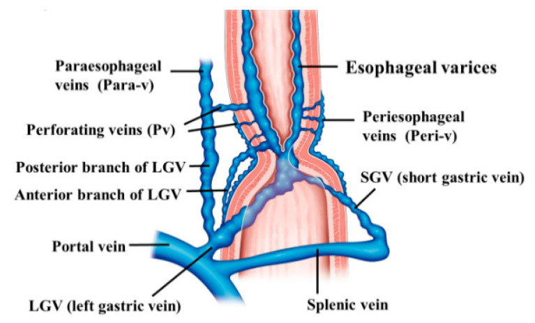
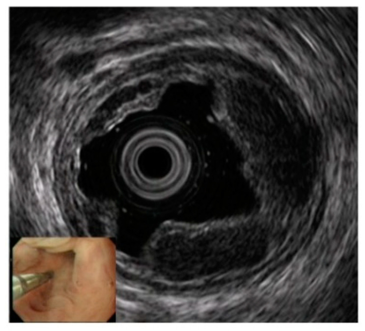
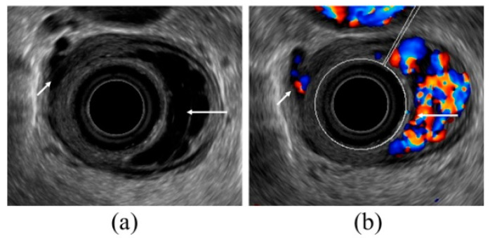
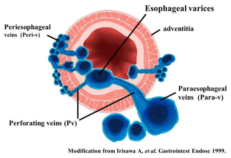
Esophageal varices are caused by the development of collateral circulation in the esophagus as a result of portal hypertension. It is important to administer appropriate preventive treatment because bleeding varices can be fatal. Esophageal varices have complex and diverse hemodynamics, and there are various variations for each case. Endoscopic ultrasound (EUS) can estimate the hemodynamics of each case. Therefore, observation by EUS in esophageal varices provides useful information, such as safe and effective treatment selection, prediction of recurrence, and appropriate follow-up after treatment. Although treatment for the esophagogastric varices can be performed without EUS imaging, understanding the local hemodynamics of the varices using EUS prior to treatment will lead to more safe and effective treatment. EUS observation is an indispensable tool for thorough variceal care.
Keywords: endoscopic injection sclerotherapy; endoscopic ultrasound; endoscopic variceal ligation; esophageal varices; hemodynamics.
Conflict of interest statement
The authors declare no conflict of interest.
Figures
Similar articles
-
Usefulness of endoscopic ultrasonographic analysis of variceal hemodynamics for the treatment of esophageal varices.Fukushima J Med Sci. 2001 Dec;47(2):39-50. doi: 10.5387/fms.47.39. Fukushima J Med Sci. 2001. PMID: 11989618
-
The Clinical Role of Endoscopic Ultrasound for Management of Bleeding Esophageal Varices in Liver Cirrhosis.Case Rep Gastroenterol. 2022 May 10;16(2):295-300. doi: 10.1159/000524529. eCollection 2022 May-Aug. Case Rep Gastroenterol. 2022. PMID: 35814797 Free PMC article.
-
Endoscopic recurrence of esophageal varices is associated with the specific EUS abnormalities: severe periesophageal collateral veins and large perforating veins.Gastrointest Endosc. 2001 Jan;53(1):77-84. doi: 10.1067/mge.2001.108479. Gastrointest Endosc. 2001. PMID: 11154493
-
Utility of endoscopic ultrasound in patients with portal hypertension.World J Gastroenterol. 2014 Oct 21;20(39):14230-6. doi: 10.3748/wjg.v20.i39.14230. World J Gastroenterol. 2014. PMID: 25339809 Free PMC article. Review.
-
Treatment modalities for bleeding esophagogastric varices.J Nippon Med Sch. 2012;79(1):19-30. doi: 10.1272/jnms.79.19. J Nippon Med Sch. 2012. PMID: 22398787 Review.
Cited by
-
Role of endoscopic ultrasound in vascular interventions: Where are we now?World J Gastrointest Endosc. 2022 Jun 16;14(6):354-366. doi: 10.4253/wjge.v14.i6.354. World J Gastrointest Endosc. 2022. PMID: 35978714 Free PMC article. Review.
-
Interobserver Reliability of the Endoscopic Ultrasound Criteria for the Diagnosis of Early Chronic Pancreatitis: Comparison between the 2009 and 2019 Japanese Diagnostic Criteria.Diagnostics (Basel). 2021 Mar 3;11(3):431. doi: 10.3390/diagnostics11030431. Diagnostics (Basel). 2021. PMID: 33802623 Free PMC article.
-
Diagnostic Accuracy of Portal Vein Flow Velocity for Esophageal Varices in Cirrhotic Patients.Cureus. 2023 Aug 16;15(8):e43592. doi: 10.7759/cureus.43592. eCollection 2023 Aug. Cureus. 2023. PMID: 37727188 Free PMC article.
-
Management of non-variceal upper gastrointestinal bleeding: role of endoscopic ultrasound-guided treatments.Therap Adv Gastroenterol. 2022 Jan 30;15:17562848211056148. doi: 10.1177/17562848211056148. eCollection 2022. Therap Adv Gastroenterol. 2022. PMID: 35126666 Free PMC article. Review.
-
Simultaneous presentation and resection of esophageal cancer and metastasis to the pancreas: Α case report and literature review.Mol Clin Oncol. 2023 Nov 17;20(1):2. doi: 10.3892/mco.2023.2700. eCollection 2024 Jan. Mol Clin Oncol. 2023. PMID: 38223405 Free PMC article.
References
Publication types
LinkOut - more resources
Full Text Sources
