. 2020 Nov 26;10(4):246.
doi: 10.3390/jpm10040246.

Integrin-Linked Kinase Is a Novel Therapeutic Target in Ovarian Cancer

Affiliations

Integrin-Linked Kinase Is a Novel Therapeutic Target in Ovarian Cancer

Michael A Ulm et al. J Pers Med. .

Abstract

Objective: The objective of this study is to identify and validate novel therapeutic target(s) in ovarian cancer.

Background: Development of targeted therapeutics in ovarian cancer has been limited by molecular heterogeneity. Although gene expression datasets are available, most of them lack appropriate pair-matched controls to define the alterations that result in the transformation of normal ovarian cells to cancerous cells.

Methods: We used microarray to compare the gene expression of treatment-naïve ovarian cancer tissue samples to pair-matched normal adjacent ovarian tissue from 24 patients. Ingenuity Pathway Analysis (IPA) was used to identify target pathways for further analysis. Integrin-linked kinase (ILK) expression in SKOV3 and OV90 cells was determined using Western blot. ILK was knocked down using CRISPR/Cas9 constructs. Subcutaneous xenograft study to determine the effect of ILK knockdown on tumor growth was performed in NOD SCID gamma mice.

Results: Significant upregulation of the ILK pathway was identified in 22 of the 24 cancer specimens, identifying it as a potential player that could contribute to the transformation of normal ovarian cells to cancerous cells. Knockdown of ILK in SKOV3 cells resulted in decreased cell proliferation and tumor growth, and inhibition of downstream kinase, AKT (protein kinase B). These results were further validated using an ILK-1 chemical inhibitor, compound 22.

Conclusion: Our initial findings validate ILK as a potential therapeutic target for molecular inhibition in ovarian cancer, which warrants further investigation.

Keywords: gene expression; integrin-linked kinase (ILK); microarray; ovarian cancer; sgRNA; xenograft.


Conflict of interest statement

No authors have any relevant conflict of interest to disclose.

Figures

Figure 1
Figure 1
Experiment design. Ovarian cancer specimens (mostly histological grade 3) and adjacent normal tissue specimens (n = 24) were used in gene expression microarray experiments and pathway analyses.
Figure 2
Figure 2
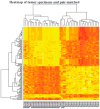
Pathway analysis. Heatmap of differentially regulated genes between tumor specimens and their respective pair-matched control specimens.
Figure 3
Figure 3
Pathway analysis of the differentially expressed genes with samples segregated based on survival. The numbers in the figures represent the number of genes (A) and the enriched pathways (BD). The different pathways are represented in each Venn circle. Survival was defined as those patients who survived less than 30 months post-diagnosis (early), more than 30 months (late), and were alive at the time of data collection (alive).
Figure 4
Figure 4
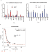
Validation of microarray data by real-time PCR. (A) Real-time PCR validation of a subset of genes identified by microarray (n = 5/gene). (B) Kaplan–Meier plot of the integrin-linked pathway (ILK-1) in the cancer genome atlas (TCGA).
Figure 5
Figure 5
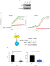
In vitro validation of the ILK pathway. (A) Protein expression of ILK-1 in two ovarian cancer cell lines, SKOV3 and OV90 and non-cancerous COS7 cells. Protein was extracted from the cells, fractionated on an SDS-PAGE, and Western blot for ILK-1 and GAPDH was performed. (B) INCUCYTE proliferation assay of SKOV3 and OV90 cell lines in the presence of vehicle (DMSO) or 3 or 10 μM ILK inhibitor compound 22. Images were obtained periodically for the indicated time-points. (C) Phosphorylation of AKT was inhibited by ILK-1 inhibitor compound 22. SKOV3 and OV90 cells were treated with compound 22 for 4 h. Cells were harvested, protein extracted, and Western blot with phospho-AKT and total AKT antibodies was performed. (D) ILK-1 siRNA inhibits SKOV3 cell proliferation. SKOV3 cells were transfected with ILK-1 or GAPDH siRNA. Six days after transfection (re-transfected after day 3) mRNA expression of ILK-1 and 18S (left) and cell proliferation (right) by sulforhodamine B (SRB) assay were measured (n = 3). ** p < 0.01; *** p < 0.001.
Figure 6
Figure 6
ILK-1 is important for SKOV3 tumor growth. (A) ILK-1 was knocked out using three CRISPR/Cas9 sgRNA. Western blot shows the knockout in SKOV3 cells. Control sgRNA was used in the vector-infected group. (B) Control and ILK-1 sgRNA knockdown cells (virus 1) were implanted in NSG mice (n = 15/group). Tumor uptake and growth were measured over the course of this study. Animals were sacrificed at the end of this study, tumors were isolated, and stored for further analysis. (C) Western blot in the tumors from animals described in panel B is provided. V1 corresponds to virus 1 (clone 1) of the three clones that were screened. (D) RNA was isolated from the tumors and the expression of genes in the vector or ILK-1 knockdown tumors (n = 3/group) was measured by microarray. (E) Representative H&E and ki67 staining of tumor sections. Statistically different genes between the two groups are represented as heatmap. ** p < 0.01.
Figure 6
Figure 6
ILK-1 is important for SKOV3 tumor growth. (A) ILK-1 was knocked out using three CRISPR/Cas9 sgRNA. Western blot shows the knockout in SKOV3 cells. Control sgRNA was used in the vector-infected group. (B) Control and ILK-1 sgRNA knockdown cells (virus 1) were implanted in NSG mice (n = 15/group). Tumor uptake and growth were measured over the course of this study. Animals were sacrificed at the end of this study, tumors were isolated, and stored for further analysis. (C) Western blot in the tumors from animals described in panel B is provided. V1 corresponds to virus 1 (clone 1) of the three clones that were screened. (D) RNA was isolated from the tumors and the expression of genes in the vector or ILK-1 knockdown tumors (n = 3/group) was measured by microarray. (E) Representative H&E and ki67 staining of tumor sections. Statistically different genes between the two groups are represented as heatmap. ** p < 0.01.

Similar articles

Cited by

References

    1. Lengyel E. Ovarian Cancer Development and Metastasis. Am. J. Pathol. 2010;177:1053–1064. doi: 10.2353/ajpath.2010.100105. - DOI - PMC - PubMed
    1. Raspagliesi F., Ditto A., Martinelli F., Haeusler E.A., Lorusso D. Advanced ovarian cancer: Omental bursa, lesser omentum, celiac, portal and triad nodes spread as cause of inaccurate evaluation of residual tumor. Gynecol. Oncol. 2013;129:92–96. doi: 10.1016/j.ygyno.2013.01.024. - DOI - PubMed
    1. Aharoni D., Meiri I., Atzmon R., Vlodavsky I., Amsterdam A. Differential effect of components of the extracellular matrix on differentiation and apoptosis. Curr. Biol. 1997;7:43–51. doi: 10.1016/S0960-9822(06)00026-1. - DOI - PubMed
    1. Sallinen H., Janhonen S., Pölönen P., Niskanen H., Liu O.H., Kivelä A., Hartikainen J.M., Anttila M., Heinäniemi M., Yla-Herttuala S., et al. Comparative transcriptome analysis of matched primary and distant metastatic ovarian carcinoma. BMC Cancer. 2019;19:1–11. doi: 10.1186/s12885-019-6339-0. - DOI - PMC - PubMed
    1. Ye X. Confluence analysis of multiple omics on platinum resistance of ovarian cancer. Eur. J. Gynaecol. Oncol. 2015;36 - PubMed

LinkOut - more resources