. 2020 Nov 26;12(12):3528.
doi: 10.3390/cancers12123528.

Epigenetic Inactivation of the Tumor Suppressor IRX1 Occurs Frequently in Lung Adenocarcinoma and Its Silencing Is Associated with Impaired Prognosis

Affiliations

Epigenetic Inactivation of the Tumor Suppressor IRX1 Occurs Frequently in Lung Adenocarcinoma and Its Silencing Is Associated with Impaired Prognosis

Miriam M Küster et al. Cancers (Basel). .

Abstract

Iroquois homeobox (IRX) encodes members of homeodomain containing genes which are involved in development and differentiation. Since it has been reported that the IRX1 gene is localized in a lung cancer susceptibility locus, the epigenetic regulation and function of IRX1 was investigated in lung carcinogenesis. We observed frequent hypermethylation of the IRX1 promoter in non-small cell lung cancer (NSCLC) compared to small cell lung cancer (SCLC). Aberrant IRX1 methylation was significantly correlated with reduced IRX1 expression. In normal lung samples, the IRX1 promoter showed lower median DNA methylation levels (<10%) compared to primary adenocarcinoma (ADC, 22%) and squamous cell carcinoma (SQCC, 14%). A significant hypermethylation and downregulation of IRX1 was detected in ADC and SQCC compared to matching normal lung samples (p < 0.0001). Low IRX1 expression was significantly correlated with impaired prognosis of ADC patients (p = 0.001). Reduced survival probability was also associated with higher IRX1 promoter methylation (p = 0.02). Inhibition of DNA methyltransferase (DNMT) activity reactivated IRX1 expression in human lung cancer cell lines. Induced DNMT3A and EZH2 expression was correlated with downregulation of IRX1. On the cellular level, IRX1 exhibits nuclear localization and expression of IRX1 induced fragmented nuclei in cancer cells. Localization of IRX1 and induction of aberrant nuclei were dependent on the presence of the homeobox of IRX1. By data mining, we showed that IRX1 is negatively correlated with oncogenic pathways and IRX1 expression induces the proapoptotic regulator BAX. In conclusion, we report that IRX1 expression is significantly associated with improved survival probability of ADC patients. IRX1 hypermethylation may serve as molecular biomarker for ADC diagnosis and prognosis. Our data suggest that IRX1 acts as an epigenetically regulated tumor suppressor in the pathogenesis of lung cancer.

Keywords: DNA methylation; IRX; adenocarcinoma; apoptosis; epigenetic; homeobox; lung cancer; tumor suppressor.


Conflict of interest statement

The authors declare no conflict of interest. The funders had no role in the design of the study; in the collection, analyses, or interpretation of data; in the writing of the manuscript or in the decision to publish the results.

Figures

Figure 1
Figure 1
Structure of IRX1 and its methylation analysis in lung cancer. (a) The IRX1 gene encodes a polypeptide of 480 aa and the protein contains a homeobox at position 145–184, a HARE-HTH (HB1, ASXL, restriction endonuclease helix-turn-helix) domain at position 188–247 and the Iroquois (IRO) box at position 313–326 (NCBI tool for conserved domain search) [26] (b) IRX1 is located on chromosome 5p15.33 in an 8.6 kb CpG island (dark green box) flanked by the two indicated CTCF binding sites (5′- and 3′-CTCFBS) [27]. (c) Overview of the analyzed 675 bp sequence located in the proximal IRX1 promoter region. The transcriptional start site (TSS; arrow) and individual CpG sites (black lines) are depicted by Python vs. CoBRA [28]. For methylation analysis, two TaqI sites of CoBRA, nine CpGs sites (1, 2, 3, 4, 5, 6, 7, 8, and 9) of bisulfite pyrosequencing, and three CpG sites (cg05534710, cg06689918, and cg09232937) of 450K array are marked. Positions of guide#1 and guide#2 for promoter deletion are marked as blue boxes upstream of the IRX1 TSS. (d) A 272 bp fragment of the IRX1 promoter was analyzed by CoBRA in five NSCLC (A549, A427, HCC15, H322, and H358), 11 SCLC (HTB171, HTB175, SCLC21H, SCLC22H, SCLC24H, H82, HTB187, H209, H510, H1092, and H1184), HeLa and in vitro methylated DNA (ivm). PCR products were mock (−) or TaqI (+) digested and analyzed on 2% agarose gels together with a 100 bp ladder. Unmethylated (u), partially methylated (pm), and methylated (m) samples are indicated.
Figure 2
Figure 2
Methylation and expression of IRX1 in cancer cell lines. (a) Methylation level of the IRX1 promoter at the three CpG sites (cg05534710, cg06689918, and cg09232937) were analyzed by Infinium Human Methylation 450 BeadChip (ilmnhm450K array) through the R2 Genomics Analysis and Visualization Platform [30]. Methylation of 244 normal lung samples (GSE52401) were compared to methylation of 1028 cancer cell lines (GSE68379). IRX1 methylation levels are plotted in beta-value (1 = 100% methylation) and p-value was calculated by one-way ANOVA. (b) Correlation analysis of IRX1 promoter methylation (beta-value) and IRX1 expression (2log ps). Methylation level of the three CpG sites of cancer cell lines (GSE68379) was plotted for IRX1 expression (11725275_at) in the corresponding mRNA 1017 of cancer cell lines [31]. Significance of correlation was calculated by the R2 platform. p < 0.05 was considered significant.
Figure 3
Figure 3
Epigenetic inactivation of IRX1 in primary lung cancers compared to matching lung. (a) Methylation level of the IRX1 promoter at the nine CpG sites was analyzed in 100 adenocarcinoma (ADC), 100 squamous cell carcinoma (SQCC), and 41 small cell lung cancer (SCLC) samples by bisulfite pyrosequencing. Average CpG methylation levels are depicted by box plot. (b) IRX1 methylation of tumors and corresponding matching lung samples were revealed by pyrosequencing. Average methylation was compared to tumor tissue by box plot and p-value was calculated. (c) Relative IRX1 expression in primary tumors and corresponding lung samples. mRNA levels were analyzed by qRTPCR normalized to the housekeeping genes ESD and RPS18 (ΔCt). Note that a higher ΔCt value indicates lower IRX1 expression. Median expression levels of IRX1 of NSCLC, ADC, and SQCC and corresponding lung samples are shown and significances (p-values) were calculated. p < 0.05 was considered significant.
Figure 4
Figure 4
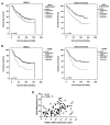
Impaired survival probability of ADC patients is associated epigenetic silencing of IRX1. (a) To correlate high and low IRX1 expression with survival probabilities, we performed the Kaplan–Meier estimator for all patients with non-small cell lung cancer (NSCLC) (n = 416) and adenocarcinoma (ADC) (n = 226) shown in months. (b). Survival probability of NSCLC and ADC patients (n = 189 and 91, respectively) was correlated with IRX1 methylation level at CpG site 8 (CpG8). (c) Correlation analysis of CpG8 methylation and relative IRX1 expression (p < 0.001). p < 0.05 was considered significant.
Figure 5
Figure 5
IRX1 is regulated by DNA methyltransferase and p300 HAT activity. (a) Deletion (Δ) of 5′- and 3′-CTCF binding site in U251 cell by CRISPR/Cas9 system. We generated two control clones, three Δ5′-CTCFBS, four Δ3′-CTCFBS, and two Δ5′+3′-CTCFBS clones and analyzed IRX1 expression for each clone. RNA levels were determined by qRTPCR in triplicates and normalized to ACTB level. IRX1 expression of control clones was set 1 for comparison. (b) Deletion of the IRX1 promoter (ΔIRX1) by CRISPR/Cas9 system. Expression of IRX1 was analyzed in 13 ΔIRX1 clones by qRTPCR and normalized to ACTB. p-value was calculated by unpaired t-test. (c) A427 and A549 cell lines were treated with the indicated concentration of 5-Aza-2′-deoxycytidine (Aza) and trichostatin A (TSA) for 4 days and RNA was isolated. IRX1 mRNA levels were analyzed in technical triplicates and normalized to ACTB. Then, 0 µM Aza treatment was set 1. (d) p300 HAT induced expression of endogenous IRX1. HeLa cells were transfected with IRX1 guide RNA constructs and p300-dCas9 or pcDNA-dCas (control). IRX1 expression was analyzed by qRTPCR after 1 µM Aza and 0.3 µM TSA treatment for 72 h and normalized to ACTB levels. (e,f) Correlation analysis of IRX1 and (e) DNMT3A or (f) EZH2 expression in primary NSCLC samples (GSE33532 data set, n = 100). Analysis was performed by R2 [30]. (g) Impaired survival probability of adenocarcinoma patients is associated with high EZH2 expression. Survival probability of ADC patients (n = 719) was correlated with EZH2 expression (low and high) through the Kaplan–Meier plotter [33]. p < 0.05 was considered significant.
Figure 6
Figure 6
Nuclear localization of IRX1 and irregular shape of IRX1 expressing nuclei. IRX1 was fused to EYFP and transfected in A549 (a) HeLa (b) and HEK293T (c) cells. Localization of IRX1 in the nucleus was analyzed by DAPI co-staining and fluorescent microscopy. IRX1 deletion construct of homeobox (ΔHomeo), HARE-HTH domain (ΔHare), and IRO box (ΔIro) were generated and transfected. The nuclear shape of transfected cells was analyzed after 24 h in A549 (80–160 cells analyzed), HeLa (200–350 cells), and HEK293 (150–300 cells). Normal nuclei exhibited a round shape and fragmented nuclei showed an irregular, lobed shape. Significance was calculated by Fisher’s exact test. White bar represents the length standard of 5 µM.
Figure 7
Figure 7
IRX1 expression is associated with expression of the apoptotic regulator BAX. (a) IRX1 wt and IRX1 deletion construct of homeobox (ΔHomeo), HARE-HTH domain (ΔHare), and IRO box (ΔIro) were transfected in HeLa cells. RNA was isolated after 48 h and BAX mRNA levels were analyzed in technical triplicates and normalized to ACTB. BAX expression was plotted relative to EYFP-control transfected HeLa cells (set = 1). (b) BAX expression values (208478_s_at) were analyzed in pcDNA3.1-GFP (control) and pcDNA3.1-IRX1-GFP transfected HEK293T cells in the data set GSE75376 by GEO2R [46,47]. (c) Expression of BAX (NM_004324) was analyzed in 143B cells after IRX1 (shIRX1) and control (shCtrl) knock down in technical triplicates in the data set GSE56255 by GEO2R [47,48].

Similar articles

Cited by

References

    1. Ferlay J., Soerjomataram I., Dikshit R., Eser S., Mathers C., Rebelo M., Parkin D.M., Forman D., Bray F. Cancer incidence and mortality worldwide: Sources, methods and major patterns in GLOBOCAN 2012. Int. J. Cancer. 2015;136:E359–E386. doi: 10.1002/ijc.29210. - DOI - PubMed
    1. Dammann R., Li C., Yoon J.H., Chin P.L., Bates S., Pfeifer G.P. Epigenetic inactivation of a RAS association domain family protein from the lung tumour suppressor locus 3p21.3. Nat. Genet. 2000;25:315–319. doi: 10.1038/77083. - DOI - PubMed
    1. Kiehl S., Zimmermann T., Savai R., Pullamsetti S.S., Seeger W., Bartkuhn M., Dammann R.H. Epigenetic silencing of downstream genes mediated by tandem orientation in lung cancer. Sci. Rep. 2017;7:3896. doi: 10.1038/s41598-017-04248-w. - DOI - PMC - PubMed
    1. Richter A.M., Kiehl S., Koger N., Breuer J., Stiewe T., Dammann R.H. ZAR1 is a novel epigenetically inactivated tumour suppressor in lung cancer. Clin. Epigenetics. 2017;9:60. doi: 10.1186/s13148-017-0360-4. - DOI - PMC - PubMed
    1. Gomez-Skarmeta J.L., Modolell J. Iroquois genes: Genomic organization and function in vertebrate neural development. Curr. Opin. Genet. Dev. 2002;12:403–408. doi: 10.1016/S0959-437X(02)00317-9. - DOI - PubMed