. 2020 Nov 26;8(12):1871.
doi: 10.3390/microorganisms8121871.

Gill Mucus and Gill Mucin O-glycosylation in Healthy and Amebic Gill Disease-Affected Atlantic Salmon

Affiliations

Gill Mucus and Gill Mucin O-glycosylation in Healthy and Amebic Gill Disease-Affected Atlantic Salmon

John Benktander et al. Microorganisms. .

Abstract

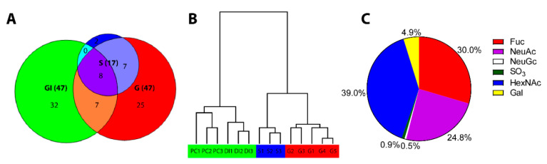
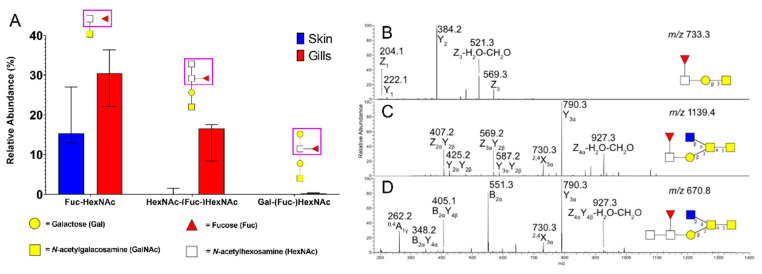
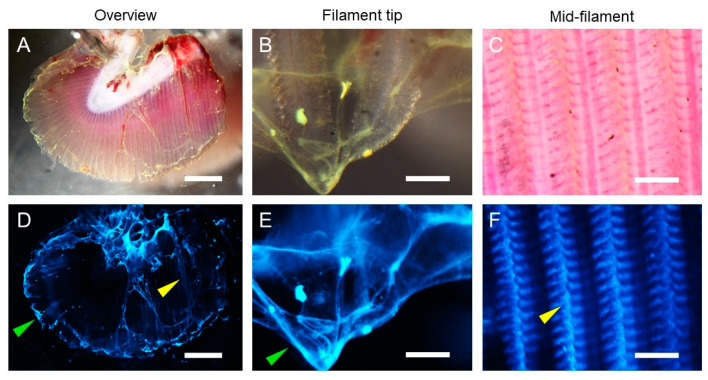
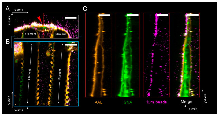
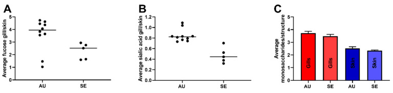
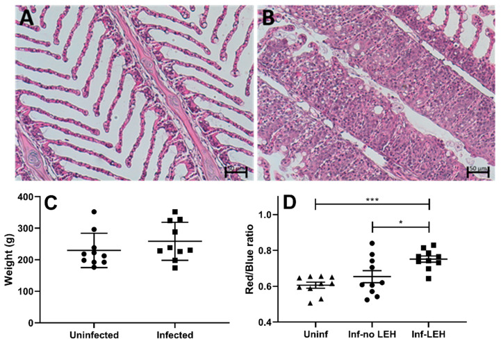
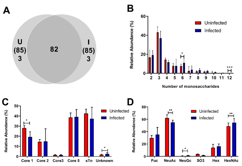
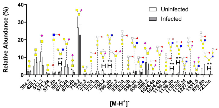
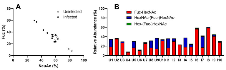
Amoebic gill disease (AGD) causes poor performance and death in salmonids. Mucins are mainly comprised by carbohydrates and are main components of the mucus covering the gill. Since glycans regulate pathogen binding and growth, glycosylation changes may affect susceptibility to primary and secondary infections. We investigated gill mucin O-glycosylation from Atlantic salmon with and without AGD using liquid chromatography-mass spectrometry. Gill mucin glycans were larger and more complex, diverse and fucosylated than skin mucins. Confocal microscopy revealed that fucosylated mucus coated sialylated mucus strands in ex vivo gill mucus. Terminal HexNAcs were more abundant among O-glycans from AGD-affected Atlantic salmon, whereas core 1 structures and structures with acidic moieties such as N-acetylneuraminic acid (NeuAc) and sulfate groups were less abundant compared to non-infected fish. The fucosylated and NeuAc-containing O-glycans were inversely proportional, with infected fish on the lower scale of NeuAc abundance and high on fucosylated structures. The fucosylated epitopes were of three types: Fuc-HexNAc-R, Gal-[Fuc-]HexNAc-R and HexNAc-[Fuc-]HexNAc-R. These blood group-like structures could be an avenue to diversify the glycan repertoire to limit infection in the exposed gills. Furthermore, care must be taken when using skin mucus as proxy for gill mucus, as gill mucins are distinctly different from skin mucins.

Keywords: Atlantic salmon; Neoparamoeba perurans; amebic gill disease; gill; glycosylation; mucin; mucosal immunology; mucus; parasite.


Conflict of interest statement

The authors declare no conflict of interest.

Figures

Figure 1
Figure 1
Characterization of Swedish gill mucin O-glycans and comparison with O-glycans from other epithelia in the same fish. (A) Venn diagram of detected O-glycan structures in skin (S), gill (G) and gastrointestinal (GI) mucins. (B) Tree view of hierarchical clustering of O-glycan profiles from pyloric caeca (PC), distal intestine (DI) and skin (S). The detailed characterization of the glycans from non-gill epithelia has been published previously [3]. (C) Pie chart of the percentage of terminal ends occupied by respective moiety among gill mucins.
Figure 2
Figure 2
Description, identification and characterization of fucosylated structures. (A) Comparison of fucosylated structures in ATS-SE (Swedish ATS) skin and gills. Bars display median with interquartile range. (B) m/z 733.3, retention time (rt): 13.1 min, (C) m/z 1139.4, rt: 15.0 min, (D) m/z 970.8 ([M-2H+]2−), rt: 16.0 min.
Figure 3
Figure 3
Ex vivo imaging of 0.2 µm fluorescent beads trapped in gill mucus. Representative micrographs showing gill overview (A,D) and higher magnification images from the gill filament tip (B,E) and mid-filament regions (C,F) from ATS-SE. Beads trapped in mucus bundles at the gill filament tip (green arrows) and between the gill filaments (yellow arrows) are indicated. Upper panels (AC) show brightfield images and equivalent fluorescence images (DF) are shown in lower panels. Images are representative of n = 5 animals; scale bars are 1 mm (A,D) and 100 µm (B,C,E,F).
Figure 4
Figure 4
Ex vivo imaging of gill mucus glycosylation and barrier properties. Representative micrographs showing confocal z-stack cross sections of ATS-SE gills stained using fluorophore-conjugated AAL (Aleuria aurantia lectin, detects fucose, orange) and SNA (Sambucus nigra, detects sialic acids, green) lectins, and overlaid with bacteria-sized 1 µm fluorescent beads (purple). (A) x/z-axis cross-section through mucus bundle (red arrow) positioned between two gill filaments. (B) x/y-axis cross-section through gill filaments shown in (A). (C) y/z-axis cross-sections through mucus bundle indicated by red dashed line in (A); panels show individual and merged fluorescent signals. Images are representative of n = 5 animals; scale bars are 100 µm.
Figure 5
Figure 5
Gill O-glycans were more fucosylated, less sialylated and larger compared to skin O-glycans, regardless of origin. (A) Fold difference in average number of fucose residues per glycan in gill compared to skin mucin O-glycans in Australian (AU) and Swedish (SE) ATS. ATS-AU skin mucin O-glycans have been described in detail previously [3]. Bars show the median. (B) Fold difference in average number of NeuAc (N-acetylneuraminic acid) residues per glycan in gill compared to skin mucin O-glycans. Bars show the median. (C) The average number of monosaccharide residues per glycan, Mean + standard error of the mean (SEM).
Figure 6
Figure 6
Disease parameters and histological characteristics in uninfected and N. perurans-infected gills. (A) Gill histology of uninfected ATS-AU showing normal filament and lamellae structure. (B) Gill histology of AGD-affected ATS demonstrates extensive hyperplasia of epithelium. N. perurans trophozoites are clearly visible on the margins between hyperplastic lesions. (C) Weight of uninfected and N.perurans-infected fish. (D) Red/blue ratio of PAS/AB secreted mucus and goblet cells of ATS. N. perurans-infected ATS gills had a tendency for more pink staining in healthy-looking areas (no LEH) as compared to that of uninfected fish (p = ns). The pink staining was significantly stronger in LEH regions of infected fish compared to both uninfected fish (p ≤ 0.001; n = 20) and healthy-looking regions of the same infected fish (“Infected, no LEH”; p ≤ 0.05; n = 20). Statistics: one-way analysis of variance (ANOVA) with Dunnett´s post hoc test against the uninfected group displayed by mean + SEM; * p ≤ 0.05; *** p ≤ 0.001. Abbreviations: LEH = lamellar epithelial hypertrophy, uninf = uninfected, inf = infected.
Figure 7
Figure 7
Overview of O-glycan characteristics in uninfected and N. perurans-infected gills. (A) Venn diagram of number of identified structures in the uninfected (U) and AGD-affected (I) groups. (B) Size distribution of the glycans, i.e., the number of monosaccharides in the structures. (C) Distribution of core structures. (D) Relative abundance of structures with corresponding terminal structures. Bars show the median with interquartile range and the significance was calculated using the Mann–Whitney U-test. *p < 0.05, **p < 0.01, ***p < 0.001.
Figure 8
Figure 8
Comparison of the 30 most abundant O-glycan structures from uninfected and N. perurans-infected gills. The figure displays the median with interquartile range and significances were calculated with the Mann–Whitney U test. * p < 0.05, **p < 0.01. Symbols: red triangle = Fuc, yellow circle = Gal, blue square = GlcNAc, yellow square = GalNAc, white square = HexNAc, purple diamond shape = NeuAc and S = sulfate group.
Figure 9
Figure 9
Fucosylated structures and their expression in N. perurans-infected and uninfected fish. (A) The relative abundance of fucosylated and sialylated structures in individual fish were plotted for comparison. The levels of fucosylated and sialylated structures are inversely proportional. (B) Graph displays the fucosylated structures separated into three groups based on terminal epitope. Seven individuals express substantial levels of HexNAc-(Fuc-)HexNAc epitopes (13–24%), while another two a moderate amount (~5%). U = uninfected, I = infected.

References

    1. Quintana-Hayashi M.P., Padra M., Padra J.T., Benktander J., Linden S.K. Mucus-Pathogen Interactions in the Gastrointestinal Tract of Farmed Animals. Microorganisms. 2018;6:55. doi: 10.3390/microorganisms6020055. - DOI - PMC - PubMed
    1. McGuckin M.A., Linden S.K., Sutton P., Florin T.H. Mucin dynamics and enteric pathogens. Nat. Rev. Microbiol. 2011;9:265–278. doi: 10.1038/nrmicro2538. - DOI - PubMed
    1. Benktander J., Venkatakrishnan V., Padra J.T., Sundh H., Sundell K., Murughan A.V.M., Maynard B., Linden S.K. Effects of size and geographical origin on Atlantic salmon, Salmo salar, mucin O-glycan repertoire. Mol. Cell. Proteom. 2019;18:1183–1196. doi: 10.1074/mcp.RA119.001319. - DOI - PMC - PubMed
    1. Jin C., Padra J.T., Sundell K., Sundh H., Karlsson N.G., Linden S.K. Atlantic Salmon Carries a Range of Novel O-Glycan Structures Differentially Localized on Skin and Intestinal Mucins. J. Proteome Res. 2015;14:3239–3251. doi: 10.1021/acs.jproteome.5b00232. - DOI - PubMed
    1. Padra M., Adamczyk B., Flahou B., Erhardsson M., Chahal G., Smet A., Jin C., Thorell A., Ducatelle R., Haesebrouck F., et al. Helicobacter suis infection alters glycosylation and decreases the pathogen growth inhibiting effect and binding avidity of gastric mucins. Mucosal Immunol. 2019;12:784–794. doi: 10.1038/s41385-019-0154-4. - DOI - PubMed

LinkOut - more resources