Functionally Relevant Differences in Plasma Fatty Acid Composition and Expression of Cytotoxic and Inhibitory NK Cell Receptors between Healthy Young and Healthy Elder Adults
- PMID: 33256224
- PMCID: PMC7759996
- DOI: 10.3390/nu12123641
Functionally Relevant Differences in Plasma Fatty Acid Composition and Expression of Cytotoxic and Inhibitory NK Cell Receptors between Healthy Young and Healthy Elder Adults
Abstract
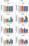
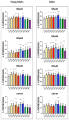
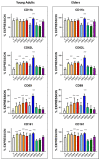
(1) Background: In the healthy ageing, NK cell number is not modified; however, their spontaneous cytotoxicity decreases. We postulated that the age-dependent decline in metabolic activities might be responsible for this effect. (2) Methods: The fatty acid profile of 30 healthy young males (23 ± 4 years old, BMI 22.1 ± 1.3) and 30 older males (63 ± 5 years old, BMI 22.9 ± 2.5) donors were evaluated along with the expression of killing (KR) and inhibitory NK receptors (KIR) at basal level and after cultivation with fatty acids for 24 h. (3) Results: Significantly higher levels of oleic (p < 0.01), arachidonic (p < 0.001), lignoceric (p < 0.001), and nervonic acids (p < 0.0001) and significantly lower levels of docosapentaenoic and docosahexaenoic acids (p < 0.01) were found in elders as compared to young adults. At basal levels, significant (p < 0.005) differences in KR and KIR expression were encountered; 12/16 antigens. Treatment of cells with saturated fatty acids or arachidonic acid (AA) significantly enhanced KR expressions (p < 0.001). AA treatment decreased inhibitory KIR expression while docosahexaenoic, and eicosapentaenoic acid increased them. (4) Conclusions: Changes in fatty acids blood levels, and KR and KIR expression in NK cell, are age-dependent. Supplementation of NK cells with eicosapentaenoic or docosahexaenoic acid enhanced inhibitory KIR receptors' expression which may improve their cell function.
Keywords: ageing; killing activating receptors; killing inhibitory receptors (KIR); natural killer cells; saturated fatty acids; unsaturated fatty acids.
Conflict of interest statement
The authors declare no conflict of interest. The funders had no role in the design of the study; in the collection, analyses, or interpretation of data; in the writing of the manuscript, or in the decision to publish the results.
Figures


Similar articles
-
Natural killer cell subsets and receptor expression in peripheral blood mononuclear cells of a healthy Korean population: Reference range, influence of age and sex, and correlation between NK cell receptors and cytotoxicity.Hum Immunol. 2017 Feb;78(2):103-112. doi: 10.1016/j.humimm.2016.11.006. Epub 2016 Nov 21. Hum Immunol. 2017. PMID: 27884732
-
Dietary supplementation with eicosapentaenoic acid, but not with other long-chain n-3 or n-6 polyunsaturated fatty acids, decreases natural killer cell activity in healthy subjects aged >55 y.Am J Clin Nutr. 2001 Mar;73(3):539-48. doi: 10.1093/ajcn/73.3.539. Am J Clin Nutr. 2001. PMID: 11237929 Clinical Trial.
-
The stage dependent changes in NK cell activity and the expression of activating and inhibitory NK cell receptors in melanoma patients.J Surg Res. 2011 Dec;171(2):637-49. doi: 10.1016/j.jss.2010.05.012. Epub 2010 Jun 8. J Surg Res. 2011. PMID: 20828749
-
Activating and inhibitory receptors of natural killer cells.Immunol Cell Biol. 2011 Feb;89(2):216-24. doi: 10.1038/icb.2010.78. Epub 2010 Jun 22. Immunol Cell Biol. 2011. PMID: 20567250 Review.
-
Regulation of self-ligands for activating natural killer cell receptors.Ann Med. 2013 Jun;45(4):384-94. doi: 10.3109/07853890.2013.792495. Epub 2013 May 23. Ann Med. 2013. PMID: 23701136 Review.
Cited by
-
Comprehensive analysis of ECHDC3 as a potential biomarker and therapeutic target for acute myeloid leukemia: Bioinformatic analysis and experimental verification.Front Oncol. 2022 Sep 12;12:947492. doi: 10.3389/fonc.2022.947492. eCollection 2022. Front Oncol. 2022. PMID: 36172164 Free PMC article.
-
Age-associated changes in innate and adaptive immunity: role of the gut microbiota.Front Immunol. 2024 Sep 16;15:1421062. doi: 10.3389/fimmu.2024.1421062. eCollection 2024. Front Immunol. 2024. PMID: 39351234 Free PMC article. Review.
-
Plasma acylcarnitines and gut-derived aromatic amino acids as sex-specific hub metabolites of the human aging metabolome.Aging Cell. 2023 Jun;22(6):e13821. doi: 10.1111/acel.13821. Epub 2023 Mar 23. Aging Cell. 2023. PMID: 36951231 Free PMC article.
References
MeSH terms
Substances
Grants and funding
LinkOut - more resources
Full Text Sources

