

Role of endothelial nitric oxide synthase for early brain injury after subarachnoid hemorrhage in mice
- PMID: 33256507
- PMCID: PMC8221759
- DOI: 10.1177/0271678X20973787
Role of endothelial nitric oxide synthase for early brain injury after subarachnoid hemorrhage in mice
Abstract
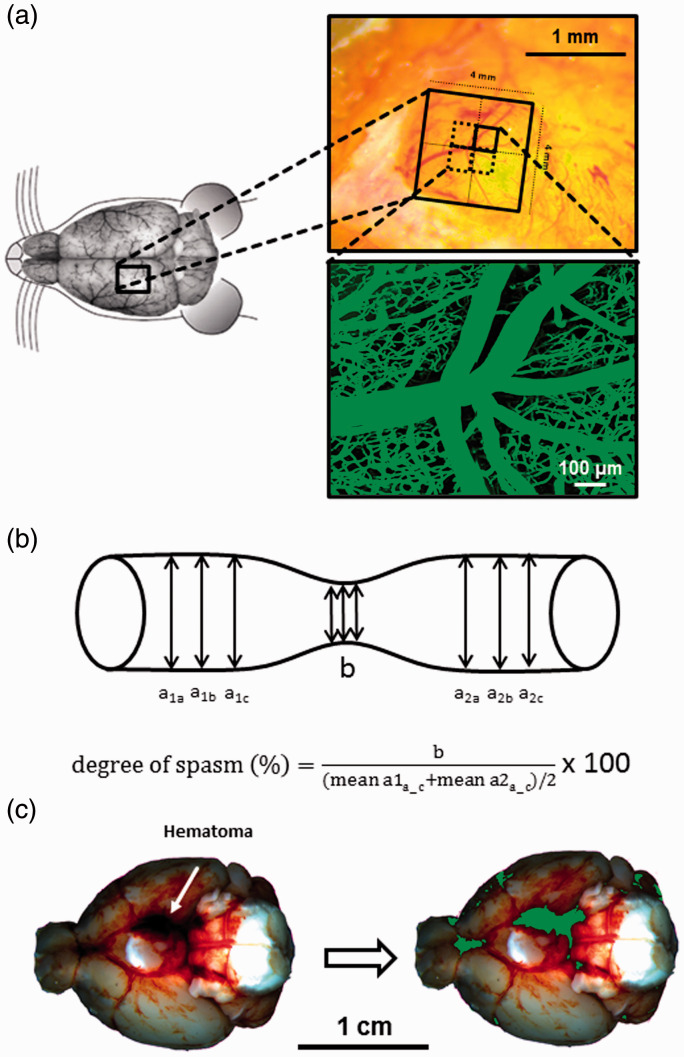
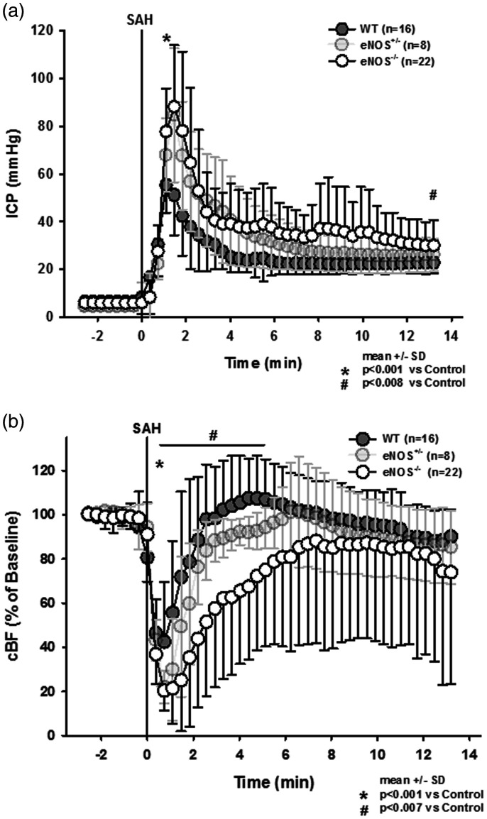
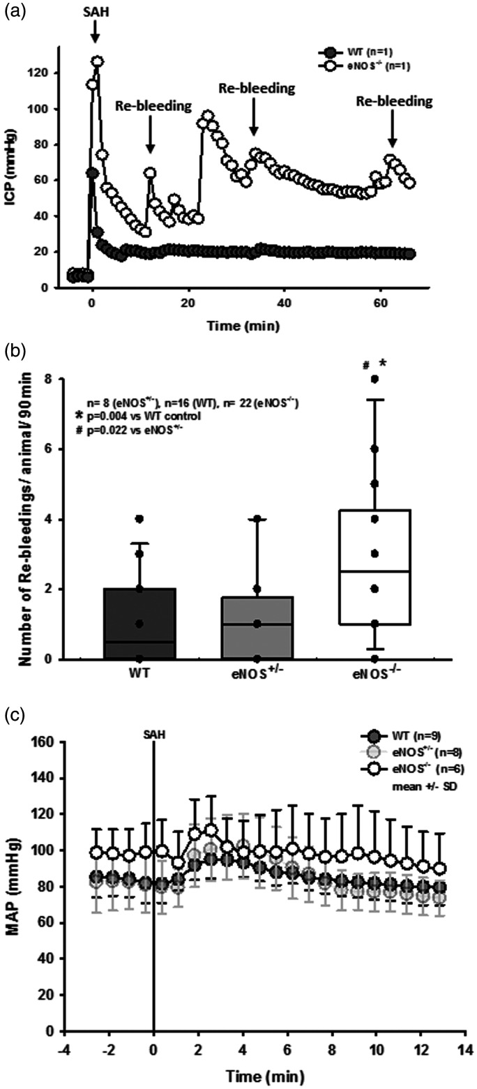
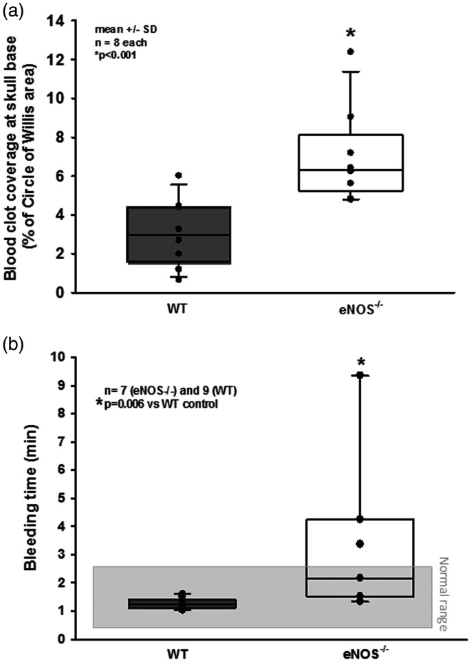
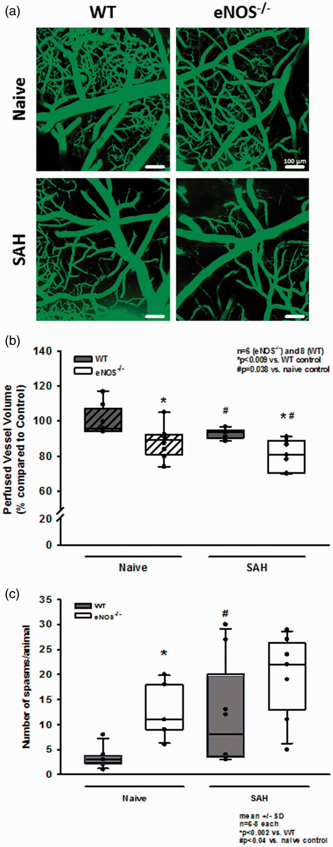
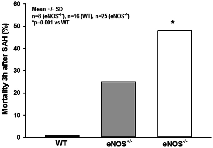
The first few hours and days after subarachnoid hemorrhage (SAH) are characterized by cerebral ischemia, spasms of pial arterioles, and a significant reduction of cerebral microperfusion, however, the mechanisms of this early microcirculatory dysfunction are still unknown. Endothelial nitric oxide production is reduced after SAH and exogenous application of NO reduces post-hemorrhagic microvasospasm. Therefore, we hypothesize that the endothelial NO-synthase (eNOS) may be involved in the formation of microvasospasms, microcirculatory dysfunction, and unfavorable outcome after SAH. SAH was induced in male eNOS deficient (eNOS-/-) mice by endovascular MCA perforation. Three hours later, the cerebral microcirculation was visualized using in vivo 2-photon-microscopy. eNOS-/- mice had more severe SAHs, more severe ischemia, three time more rebleedings, and a massively increased mortality (50 vs. 0%) as compared to wild type (WT) littermate controls. Three hours after SAH eNOS-/- mice had fewer perfused microvessels and 40% more microvasospasms than WT mice. The current study indicates that a proper function of eNOS plays a key role for a favorable outcome after SAH and helps to explain why patients suffering from hypertension or other conditions associated with impaired eNOS function, have a higher risk of unfavorable outcome after SAH.
Keywords: Subarachnoid hemorrhage; early brain injury; endothelial NOS; microvasospasm; nitric oxide.
Conflict of interest statement
Figures
Similar articles
-
Nitric oxide inhalation reduces brain damage, prevents mortality, and improves neurological outcome after subarachnoid hemorrhage by resolving early pial microvasospasms.J Cereb Blood Flow Metab. 2016 Dec;36(12):2096-2107. doi: 10.1177/0271678X15605848. Epub 2015 Nov 2. J Cereb Blood Flow Metab. 2016. PMID: 26661144 Free PMC article.
-
Role of Pial Microvasospasms and Leukocyte Plugging for Parenchymal Perfusion after Subarachnoid Hemorrhage Assessed by In Vivo Multi-Photon Microscopy.Int J Mol Sci. 2021 Aug 6;22(16):8444. doi: 10.3390/ijms22168444. Int J Mol Sci. 2021. PMID: 34445151 Free PMC article.
-
Genetic elimination of eNOS reduces secondary complications of experimental subarachnoid hemorrhage.J Cereb Blood Flow Metab. 2013 Jul;33(7):1008-14. doi: 10.1038/jcbfm.2013.49. Epub 2013 Apr 3. J Cereb Blood Flow Metab. 2013. PMID: 23549379 Free PMC article.
-
Role of nitric oxide and mechanisms involved in cerebral injury after subarachnoid hemorrhage: is nitric oxide a possible answer to cerebral vasospasm?J Neurosurg Sci. 2016 Sep;60(3):385-91. Epub 2015 Jan 20. J Neurosurg Sci. 2016. PMID: 25600552 Review.
-
Mechanisms of acute brain injury after subarachnoid hemorrhage.Neurol Res. 2006 Jun;28(4):381-98. doi: 10.1179/016164106X114991. Neurol Res. 2006. PMID: 16759442 Review.
Cited by
-
Inhaled Nitric Oxide Treatment for Aneurysmal SAH Patients With Delayed Cerebral Ischemia.Front Neurol. 2022 Feb 18;13:817072. doi: 10.3389/fneur.2022.817072. eCollection 2022. Front Neurol. 2022. PMID: 35250821 Free PMC article.
-
Inflammation and Oxidative Stress: Potential Targets for Improving Prognosis After Subarachnoid Hemorrhage.Front Cell Neurosci. 2021 Sep 24;15:739506. doi: 10.3389/fncel.2021.739506. eCollection 2021. Front Cell Neurosci. 2021. PMID: 34630043 Free PMC article. Review.
-
Early Hydrogen-Oxygen Gas Mixture Inhalation in Patients with Aneurysmal Subarachnoid Hemorrhage (HOMA): study protocol for a randomized controlled trial.Trials. 2024 Jun 11;25(1):377. doi: 10.1186/s13063-024-08231-5. Trials. 2024. PMID: 38863026 Free PMC article.
-
Adrenomedullin Is a Diagnostic and Prognostic Biomarker for Acute Intracerebral Hemorrhage.Curr Issues Mol Biol. 2021 Jun 11;43(1):324-334. doi: 10.3390/cimb43010027. Curr Issues Mol Biol. 2021. PMID: 34208106 Free PMC article.
-
Ferroptosis in early brain injury after subarachnoid hemorrhage: review of literature.Chin Neurosurg J. 2024 Feb 13;10(1):6. doi: 10.1186/s41016-024-00357-4. Chin Neurosurg J. 2024. PMID: 38347652 Free PMC article. Review.
References
-
- van Gijn J, Kerr RS, Rinkel GJ.Subarachnoid haemorrhage. Lancet 2007; 369: 306–318. - PubMed
-
- Mayer S, Kreiter K.Quality of life after subarachnoid hemorrhage. J Neurosurg 2002; 97: 741–742. - PubMed
-
- Schwartz C, Pfefferkorn T, Ebrahimi C, et al.. Long-term neurological outcome and quality of life after world federation of neurosurgical societies grades IV and V aneurysmal subarachnoid hemorrhage in an interdisciplinary treatment concept. Neurosurgery 2017; 80: 967–974. DOI: 2966486 [pii];10.1093/neuros/nyw138 [doi]. - PubMed
-
- Zijlmans JL, Coert BA, van den Berg R, et al.. Unfavorable outcome in patients with aneurysmal subarachnoid hemorrhage WFNS grade I. World Neurosurg 2018; 118: e217–e222. [pii];10.1016/j.wneu.2018.06.157 [doi]. - PubMed
-
- Cahill J, Cahill WJ, Calvert JW, et al.. Mechanisms of early brain injury after subarachnoid hemorrhage. J Cereb Blood Flow Metab 2006; 26: 1341–1353. - PubMed
Publication types
MeSH terms
Substances
LinkOut - more resources
Full Text Sources
