

Ex Vivo Imaging of Ultrasound-Stimulated Metabolic Activity in Rat Pancreatic Slices
- PMID: 33257101
- PMCID: PMC7856007
- DOI: 10.1016/j.ultrasmedbio.2020.10.021
Ex Vivo Imaging of Ultrasound-Stimulated Metabolic Activity in Rat Pancreatic Slices
Abstract
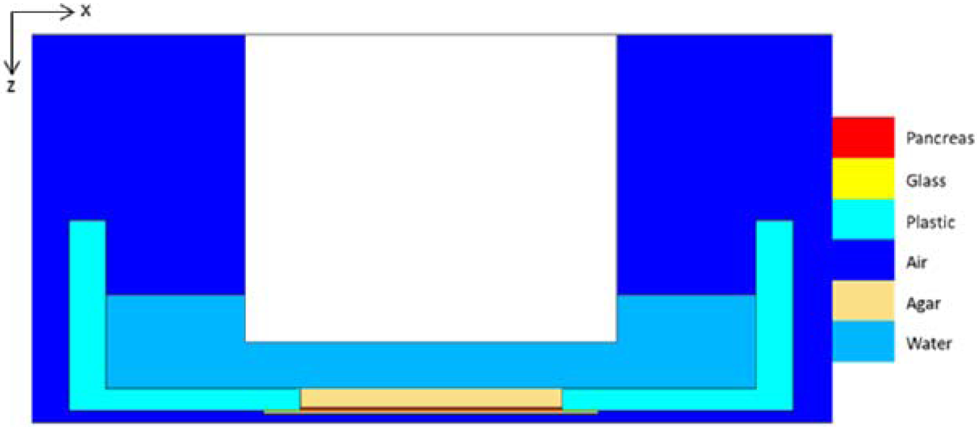
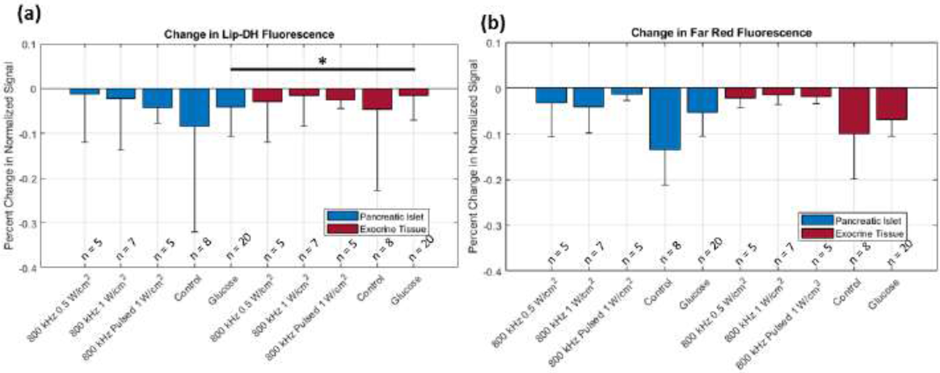
Ultrasound has previously been reported to produce a reversible stimulatory effect in cultured rat beta cells. Here, we quantified and assessed dynamic metabolic changes in an in situ pancreatic slice model evoked by ultrasound application. After plating, pancreas slices were imaged using a confocal microscope at 488 and 633 nm to image lipodamine dehydrogenase (Lip-DH) autofluorescence and a far red fluorescence, respectively. Ultrasound was applied at intensities of 0.5 and 1 W/cm2 at both 800 kHz and 1 MHz. Additionally, 800 kHz at 1 W/cm2 was applied in a pulsing scheme. No ultrasound (control) and glucose application experiments were performed. A difference in fluorescence signal before and after treatment application was the metric for analysis. Comparison of experimental groups using far red fluorescence revealed significant differences between all experimental groups and control in the islet (p < 0.05) and between all ultrasound experimental groups and control (p < 0.05) in pancreatic exocrine tissue. However, this difference in response between control and glucose did not exist in the exocrine tissue. We also observed using Lip-DH autofluorescence that glucose produces a significantly increased metabolic response in islet tissue compared with exocrine tissue (p < 0.05). Pulsed ultrasound appeared to increase metabolic activity in the pancreatic slice in a more consistent manner compared with continuous ultrasound application. Our results indicate that therapeutic ultrasound may have a stimulatory metabolic effect on the pancreatic islets similar to that of glucose.
Keywords: Fluorescence imaging; Pancreas; Therapeutic ultrasound; Type 2 diabetes.
Copyright © 2020 World Federation for Ultrasound in Medicine & Biology. Published by Elsevier Inc. All rights reserved.
Figures


Similar articles
-
Multipotential nestin-positive stem cells isolated from adult pancreatic islets differentiate ex vivo into pancreatic endocrine, exocrine, and hepatic phenotypes.Diabetes. 2001 Mar;50(3):521-33. doi: 10.2337/diabetes.50.3.521. Diabetes. 2001. PMID: 11246871
-
18F-Fallypride PET of pancreatic islets: in vitro and in vivo rodent studies.J Nucl Med. 2011 Jul;52(7):1125-32. doi: 10.2967/jnumed.111.088583. Epub 2011 Jun 16. J Nucl Med. 2011. PMID: 21680697
-
Effects of islet hormones on nerve-mediated and acetylcholine-evoked secretory responses in the isolated pancreas of normal and diabetic rats.Int J Mol Med. 1998 Mar;1(3):627-34. doi: 10.3892/ijmm.1.3.627. Int J Mol Med. 1998. PMID: 9852277
-
[Effect of Electroacupuncture at "Weiwanxiashu" (EX-B 3) on Islet Morphology and the Expression of Pancreatic Glucagon-like Peptide-1 Receptor in Type 2 Diabetes Rats].Zhen Ci Yan Jiu. 2017 Apr 25;42(2):107-13. Zhen Ci Yan Jiu. 2017. PMID: 29071956 Chinese.
-
Assessment of islet specificity of dihydrotetrabenazine radiotracer binding in rat pancreas and human pancreas.J Nucl Med. 2010 Sep;51(9):1439-46. doi: 10.2967/jnumed.109.074492. Epub 2010 Aug 18. J Nucl Med. 2010. PMID: 20720057
Cited by
-
Low-intensity therapeutic ultrasound effects on intracellular sodium and reactive oxygen species in ex vivo human islets for control of insulin release.Ultrasonics. 2025 Nov;155:107737. doi: 10.1016/j.ultras.2025.107737. Epub 2025 Jun 17. Ultrasonics. 2025. PMID: 40543425
-
Modeling of Ultrasound Stimulation of Adolescent Pancreas for Insulin Release Therapy.J Ultrasound Med. 2023 Aug;42(8):1699-1707. doi: 10.1002/jum.16189. Epub 2023 Feb 1. J Ultrasound Med. 2023. PMID: 36723381 Free PMC article.
References
-
- Abdollahi A, Domhan S, Jenne JW, Hallaj M, Delì Aqua G, Mueckenthaler M, Richter A, Martin H, Debus J, Ansorge W, Hynynen K, Huber PE. Apoptosis signals in lymphoblasts induced by focused ultrasound. FASEB J 2004;18(12):1413–4. - PubMed
-
- Baschong W, Suetterlin R, Hubert Laeng R. Control of autofluorescence of archival formaldehyde-fixed, paraffin-embedded tissue in confocal laser scanning microscopy (CLSM). J Histochem Cytochem Histochemical Society, 2001;49:1565–1571. - PubMed
-
- Bystritsky A, Korb AS, Douglas PK, Cohen MS, Melega WP, Mulgaonkar AP, DeSalles A, Min B-K, Yoo S-S. A review of low-intensity focused ultrasound pulsation. Brain Stimul, 2011;4:125–136. - PubMed
-
- Cade JE, Hanison J. The pancreas. Anaesth Intensive Care Med, 2017;18:527–531.
-
- CDC. Diabetes Report Card, 2014. Atlanta, GA, USA Centers Dis Control Prev US Dept Heal Hum Serv; 2015.
Publication types
MeSH terms
Grants and funding
LinkOut - more resources
Full Text Sources
Other Literature Sources
