Non-sebaceous lymphadenoma of the submandibular gland: diagnostic challenges in the head and neck cancer pathway
- PMID: 33257384
- PMCID: PMC7705567
- DOI: 10.1136/bcr-2020-238099
Non-sebaceous lymphadenoma of the submandibular gland: diagnostic challenges in the head and neck cancer pathway
Abstract
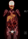
Non-sebaceous lymphadenoma (NSLA) is a rare benign salivary gland tumour with lymphoid and epithelial components and without sebaceous differentiation. The large majority of the reported cases arise within the parotid gland. We present an NSLA arising from the submandibular gland. The tumour presented as a painless longstanding neck lump. Ultrasound, fine needle aspiration, MRI and positron emission tomography found features supportive of squamous cell carcinoma. The patient was treated with surgery for oropharyngeal carcinoma of unknown origin, in accordance with local and national guidelines. The final histological assessment revealed the level Ib neck lesion to be NSLA. Although a rare occurrence, these lesions may pose a diagnostic challenge in the head and neck cancer pathway.
Keywords: otolaryngology / ENT; pathology.
© BMJ Publishing Group Limited 2020. No commercial re-use. See rights and permissions. Published by BMJ.
Conflict of interest statement
Competing interests: None declared.
Figures


Similar articles
-
Sebaceous lymphadenoma of submandibular gland.J Indian Med Assoc. 2013 Jan;111(1):62-3. J Indian Med Assoc. 2013. PMID: 24000514
-
A rare case of sebaceous lymphadenoma: Cytohistological correlation of preoperative cytomorphologic findings.Ann Diagn Pathol. 2022 Dec;61:152058. doi: 10.1016/j.anndiagpath.2022.152058. Epub 2022 Oct 29. Ann Diagn Pathol. 2022. PMID: 36334412
-
Lymphadenoma lacking sebaceous differentiation in the parotid gland.J Formos Med Assoc. 2004 Jun;103(6):459-62. J Formos Med Assoc. 2004. PMID: 15278191
-
Unilateral submandibular gland aplasia masquerading as cancer nodal metastasis.Am J Otolaryngol. 2008 Nov-Dec;29(6):432-4. doi: 10.1016/j.amjoto.2007.12.002. Epub 2008 Jun 16. Am J Otolaryngol. 2008. PMID: 19144308 Review.
-
Pitfalls in the staging of cancer of the major salivary gland neoplasms.Neuroimaging Clin N Am. 2013 Feb;23(1):107-22. doi: 10.1016/j.nic.2012.08.009. Neuroimaging Clin N Am. 2013. PMID: 23199664 Review.
References
-
- Auclair PL, Ellis GL, Gnepp DR. Other benign epithelial neoplasms : Ellis GL, Auclair PL, Gnepp DR, “Surgical pathology of the salivary glands”. Saunders, Philadelphia, PA, 1991.
-
- Barnes L, Eveson JW, Reichart P, et al. . World Health organization classification of tumours—pathology and genetics of head and neck tumours. Iarc 2005.
Publication types
MeSH terms
LinkOut - more resources
Full Text Sources
Medical
Research Materials
Miscellaneous
