Case Reports
. 2020 Dec 16;12(574):eabe4282.
doi: 10.1126/scitranslmed.abe4282. Epub 2020 Nov 30.

Lung transplantation for patients with severe COVID-19

Affiliations
Case Reports

Lung transplantation for patients with severe COVID-19

Ankit Bharat et al. Sci Transl Med. .

Abstract

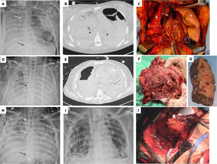
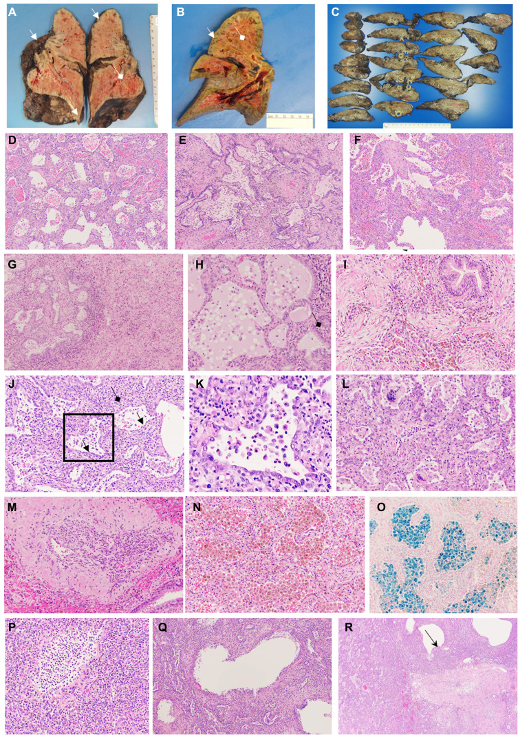
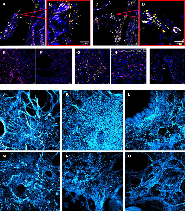
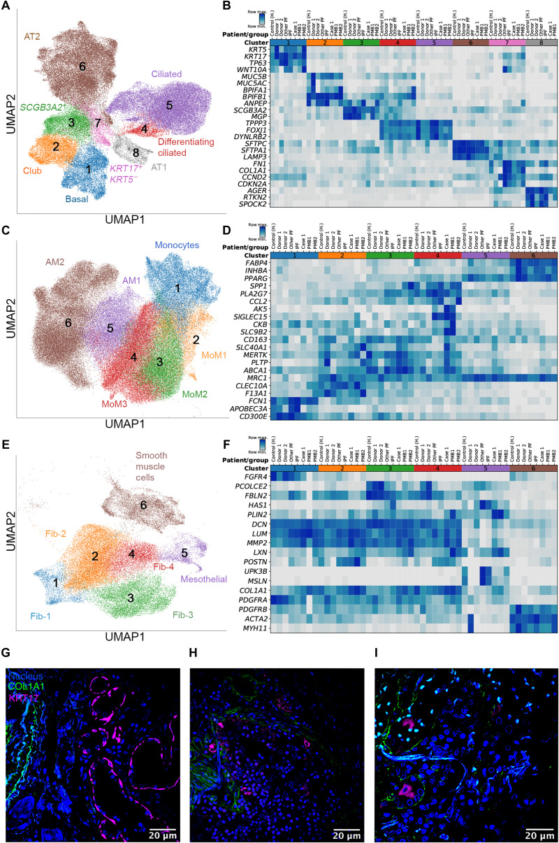
Lung transplantation can potentially be a life-saving treatment for patients with nonresolving COVID-19-associated respiratory failure. Concerns limiting lung transplantation include recurrence of SARS-CoV-2 infection in the allograft, technical challenges imposed by viral-mediated injury to the native lung, and the potential risk for allograft infection by pathogens causing ventilator-associated pneumonia in the native lung. Additionally, the native lung might recover, resulting in long-term outcomes preferable to those of transplant. Here, we report the results of lung transplantation in three patients with nonresolving COVID-19-associated respiratory failure. We performed single-molecule fluorescence in situ hybridization (smFISH) to detect both positive and negative strands of SARS-CoV-2 RNA in explanted lung tissue from the three patients and in additional control lung tissue samples. We conducted extracellular matrix imaging and single-cell RNA sequencing on explanted lung tissue from the three patients who underwent transplantation and on warm postmortem lung biopsies from two patients who had died from COVID-19-associated pneumonia. Lungs from these five patients with prolonged COVID-19 disease were free of SARS-CoV-2 as detected by smFISH, but pathology showed extensive evidence of injury and fibrosis that resembled end-stage pulmonary fibrosis. Using machine learning, we compared single-cell RNA sequencing data from the lungs of patients with late-stage COVID-19 to that from the lungs of patients with pulmonary fibrosis and identified similarities in gene expression across cell lineages. Our findings suggest that some patients with severe COVID-19 develop fibrotic lung disease for which lung transplantation is their only option for survival.


Figures

Fig. 1
Fig. 1. Radiographic and intraoperative findings in lung transplant recipients with severe COVID-19.
(A to C) Radiographic and intraoperative findings for case 1 with severe COVID-19 who underwent lung transplantation. (A) Pretransplant chest radiograph (day 38 after onset of ARDS) for case 1, revealing opacification of the right lung and a left lower lobe necrotic cavity attributed to pneumonia caused by Serratia marcascens. A tube thoracostomy was required to treat right spontaneous hemothorax and peumothoraces. In addition, the image shows ECMO cannulas (inflow cannula carrying oxygenated blood to the patient is indicated by the white arrow, and outflow cannula carrying deoxygenated blood from the patient to the ECMO machine is indicated by the black arrow). (B) Severe ARDS and lower lung lobe necrosis in case 1 were confirmed by cross-sectional computed tomography imaging. (C) Shown is an intraoperative image revealing contrasting features between the diseased native right lung (d) and the newly transplanted left lung (n). The photograph was taken immediately after the transplant of the left lung and before proceeding onto right lung transplantation. The pericardial sac (white arrow) containing the heart was opened to gain access to the aorta and place the outflow cannula of the venoarterial ECMO. (D to F) Radiographic and intraoperative findings for case 2 with severe COVID-19 who underwent lung transplantation. (D) Pretransplant chest radiograph (at day 98 after onset of ARDS) for case 2 revealing bilateral lung opacifications and a necrotic cavity in the right lung attributed to pneumonia caused by Pseudomonas aeruginosa. A chest tube to treat bronchopleural fistula is visible. The dual lumen ECMO cannula is indicated by the black arrow. (E) Computed tomography imaging indicates severe ARDS and development of a necrotic cavity in the right lung. (F) Freshly explanted right lung of case 2 with extensive pleural inflammation and loss of identifiable anatomical planes. (G) Formalin-fixed explanted right lung of case 1 demonstrating the development of lung cavities. (H to J) Radiographic and intraoperative findings for case 3 with severe COVID-19 who underwent lung transplantation. (H) Pretransplant chest radiograph (day 86 after onset of severe ARDS) for case 3, revealing extensive consolidation (lack of air) with right fibrothorax. In addition, the image shows ECMO cannulas (inflow cannula carrying oxygenated blood to the patient is indicated by the white arrow, and the outflow cannula carrying deoxygenated blood from the patient to the ECMO machine is indicated by the black arrow). (I) Posttransplant day 1 chest radiograph for case 3 demonstrating the expected appearance of new lung allografts after bilateral lung transplantation. (J) Intraoperative photograph after implantation of the left lung and explantation of the right lung revealing the right hemithorax. Diffuse pleuritis with severe thickening of parietal pleura and neoangiogenesis (white arrows) were noted as in the other two cases.
Fig. 2
Fig. 2. Common histological features of lung explants.
Shown are common histological features of lung explants from three patients with severe COVID-19 who underwent lung transplantation. (A to C) Gross pathology images of explanted lungs from case 1 (A), case 2 (B), and case 3 (C). Cystic structures are evident on the lung surface and in the lung parenchyma (white arrows) along with diffuse fibrosis. Purulent secretions in the airways suggestive of bronchopneumonia are indicated by white diamond arrows. (D to R) Remaining images show sections of the explanted lung from cases 1, 2, and 3 stained with hematoxylin and eosin, except for image (O), which was stained with Prussian blue to detect iron deposition. (D) Image shows lung alveoli in the explanted lung from case 1 demonstrating hemorrhage, interstitial fibrosis, and prominent reactive pneumocytes (100×). (E) Bronchiolitis and bronchiolar fibrosis with microscopic honeycombing were observed for the explanted lung from case 2 (200×). (F) Organizing areas of alveolar hemorrhage are visible on this image of explanted lung from case 3 (100×). (G) Microscopic honeycombing adjacent to an area of more dense fibrosis with interstitial expansion and inflammatory infiltrates is visible on this image of explanted lung from case 2 (100×). (H) Bronchiolitis and fibrosis (black diamond arrow) are visible on this image of explanted lung from case 2 (200×). (I) An area of organizing pneumonia showing whorls of fibroblasts in the airways and interstitial expansion is observed on this image of explanted lung from case 2 (200×). (J) Interstitial fibrosis and bronchiolar fibrosis (black arrows) are visible in this image of explanted lung from case 2 (100×). (K) Enlarged inset from (J) reveals a bronchiole surrounded by interstitial fibrosis and cuboidal epithelia. (L) Area of interstitial fibrosis and microscopic honeycombing with multinucleated giant cells in the airspaces is visible on this image of explanted lung from case 1 (200×). (M) A medium-size blood vessel with an organizing thrombus and recannulation can be observed in this image of explanted lung from case 2 (100×). (N) Interstitial inflammation and expansion with alveoli filled with pigmented macrophages are visible in this image of explanted lung from case 1 (100×). (O) Staining for iron revealed alveolar macrophages laden with hemosiderin (blue) in this image of explanted lung from case 1 (100×). (P) This image of explanted lung from case 1 shows formation of cystic airspaces with neutrophilic inflammatory infiltrates (200×). (Q) This image of explanted lung from case 1 reveals cystic airspaces lined by histiocytes and hyperplastic epithelia (100×), and (R) this image reveals a mature cyst (black arrow) near an area of airway fibrosis (40×).
Fig. 3
Fig. 3. smFISH and matrix imaging in cleared lung sections from patients with severe COVID-19.
(A to D) RNAScope and immunohistochemistry of lung autopsy tissue from a patient who declined interventions for COVID-19–induced respiratory failure (palliative COVID-19). Nuclear staining (blue), positive strand SARS-CoV-2 RNA (yellow), negative strand SARS-CoV-2 RNA (cyan), and CD206 (magenta). Positive strand SARS-CoV-2 RNA (yellow) was detected in cells with morphological features suggestive of epithelial cells (white arrows); negative strand SARS-CoV-2 RNA was also detected (cyan; circular arrow). (E and F) RNAScope images of the explanted lung from case 1 who underwent lung transplantation. There is yellow autofluorescence but no staining for SARS-CoV-2 positive or negative strand RNA in either left (E) or right (F) explanted lung. (G and H) RNAScope images of the explanted lung of case 2 who underwent lung transplantation, showing no evidence of SARS-CoV-2 RNA. (I) RNAScope image of a postmortem lung biopsy from a patient who died of COVID-19 (PMB1), showing no evidence of SARS-CoV-2 RNA. (J to O) Cleared lung tissue allowing visualization of the collagen structure and matrix of lung tissue (cyan); ×20 magnification. (J) Normal lung from a patient who died of pulmonary embolism. (K) An explanted lung from case 1 who underwent lung transplantation. (L and M) Shown are postmortem lung biopsies from two patients who died of COVID-19 (PMB1 and PMB2). (N and O) Shown are lung explants from two patients with idiopathic pulmonary fibrosis (IPF1 and IPF2) who underwent lung transplantation.
Fig. 4
Fig. 4. Single-cell RNA sequencing of lung tissue from patients with severe COVID-19.
(A, C, and E) Uniform Manifold Approximation and Projection (UMAP) plots showing individual populations of epithelial cells (A), macrophages (C), and mesenchymal cells (E). (B, D, and F) Heatmaps illustrating expression of select marker genes in epithelial cells (B), macrophages (D), and mesenchymal cells (F). Gene expression for the pulmonary fibrosis dataset of Habermann et al. (16) is shown as an average per condition; gene expression for the end-stage COVID-19 dataset is shown per individual patient. Labels on heatmaps (B, D, and F) correspond to the following samples: Control (H), healthy controls; IPF, idiopathic pulmonary fibrosis samples; Other PF, samples from patients with other forms of pulmonary fibrosis, all from the Habermann et al. dataset. Donor 1, Donor 2, control donor lungs; Case 1, lung transplant case 1; PMB1, PMB2, postmortem lung biopsies from two patients with COVID-19, all from the end-stage COVID-19 dataset. (G) Immunofluorescence microscopy revealed KRT17 staining (magenta) of flat epithelial cells resembling alveolar type 1 cells in nonfibrotic lung tissue from a patient who died of COVID-19 (palliative COVID-19). (H and I) Immunofluorescence microscopy revealed KRT17 staining (magenta) of distal explanted lung tissue from a patient with COVID-19 undergoing lung transplantation (H) and lung tissue from a patient (PMB1) who died from late-stage severe COVID-19 (I). Normal lung architecture is lacking, and solitary KRT17-positive cells (magenta) can be observed close to COL1A1-positive cells (green). Scale bars, 20 um.

Update of

References

    1. Geleris J., Sun Y., Platt J., Zucker J., Baldwin M., Hripcsak G., Labella A., Manson D. K., Kubin C., Barr R. G., Sobieszczyk M. E., Schluger N. W., Observational study of hydroxychloroquine in hospitalized patients with Covid-19. N. Engl. J. Med. 382, 2411–2418 (2020). - PMC - PubMed
    1. Richardson S., Hirsch J. S., Narasimhan M., Crawford J. M., Ginn T. M., Davidson K. W.; Northwell COVID- Research Consortium, Barnaby D. P., Becker L. B., Chelico J. D., Cohen S. L., Cookingham J., Coppa K., Diefenbach M. A., Dominello A. J., Duer-Hefele J., Falzon L., Gitlin J., Hajizadeh N., Harvin T. G., Hirschwerk D. A., Kim E. J., Kozel Z. M., Marrast L. M., Mogavero J. N., Osorio G. A., Qiu M., Zanos T. P., Presenting characteristics, comorbidities, and outcomes among 5700 patients hospitalized with COVID-19 in the New York City Area. JAMA 323, 2052–2059 (2020). - PMC - PubMed
    1. The RECOVERY Collaborative Group , Dexamethasone in Hospitalized Patients with Covid-19 – Preliminary Report. N. Engl. J. Med. 10.1056/NEJMoa2021436 , (2020). - PMC - PubMed
    1. McMahon J. H., Udy A., Peleg A. Y., Remdesivir for the treatment of Covid-19 – Preliminary report. N. Engl. J. Med. 383, 992–994 (2020). - PubMed
    1. van der Mark S. C., Hoek R. A. S., Hellemons M. E., Developments in lung transplantation over the past decade. Eur. Respir. Rev. 29, 190132 (2020). - PMC - PubMed

Publication types

MeSH terms