. 2021 Jan 25;203(4):e00568-20.
doi: 10.1128/JB.00568-20. Print 2021 Jan 25.

The Two TpsB-Like Proteins in Anabaena sp. Strain PCC 7120 Are Involved in Secretion of Selected Substrates

Affiliations

The Two TpsB-Like Proteins in Anabaena sp. Strain PCC 7120 Are Involved in Secretion of Selected Substrates

Giang Ngo et al. J Bacteriol. .

Abstract

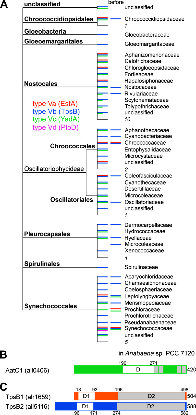
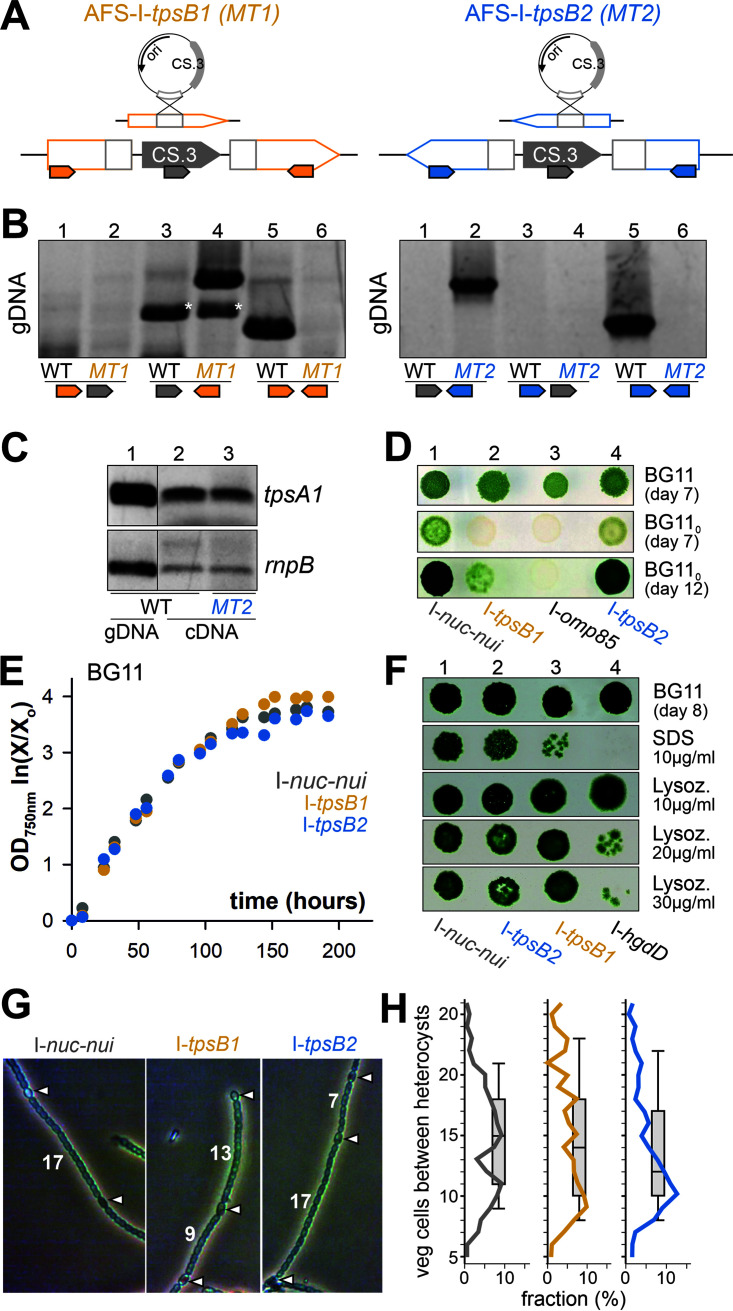
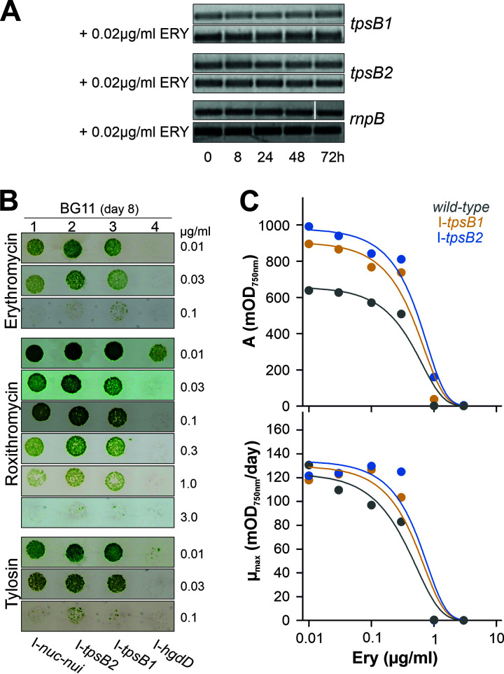
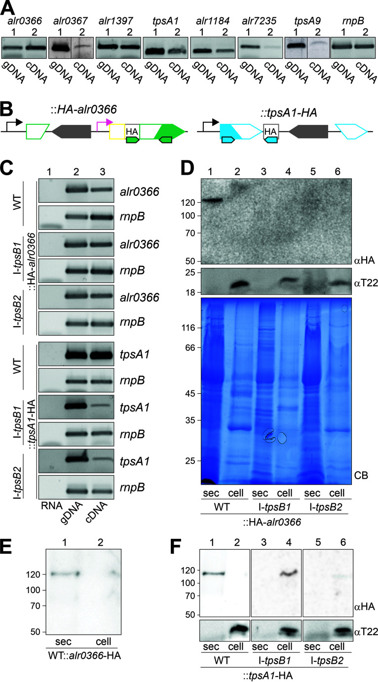
The outer membrane of Gram-negative bacteria acts as an initial diffusion barrier that shields the cell from the environment. It contains many membrane-embedded proteins required for functionality of this system. These proteins serve as solute and lipid transporters or as machines for membrane insertion or secretion of proteins. The genome of Anabaena sp. strain PCC 7120 codes for two outer membrane transporters termed TpsB1 and TpsB2. They belong to the family of the two-partner secretion system proteins which are characteristic of pathogenic bacteria. Because pathogenicity of Anabaena sp. strain PCC 7120 has not been reported, the function of these two cyanobacterial TpsB proteins was analyzed. TpsB1 is encoded by alr1659, while TpsB2 is encoded by all5116 The latter is part of a genomic region containing 11 genes encoding TpsA-like proteins. However, tpsB2 is transcribed independently of a tpsA gene cluster. Bioinformatics analysis revealed the presence of at least 22 genes in Anabaena sp. strain PCC 7120 putatively coding for substrates of the TpsB system, suggesting a rather global function of the two TpsB proteins. Insertion of a plasmid into each of the two genes resulted in altered outer membrane integrity and antibiotic resistance. In addition, the expression of genes coding for the Clp and Deg proteases is dysregulated in these mutants. Moreover, for two of the putative substrates, a dependence of the secretion on functional TpsB proteins could be confirmed. We confirm the existence of a two-partner secretion system in Anabaena sp. strain PCC 7120 and predict a large pool of putative substrates.IMPORTANCE Cyanobacteria are important organisms for the ecosystem, considering their contribution to carbon fixation and oxygen production, while at the same time some species produce compounds that are toxic to their environment. As a consequence, cyanobacterial overpopulation might negatively impact the diversity of natural communities. Thus, a detailed understanding of cyanobacterial interaction with the environment, including other organisms, is required to define their impact on ecosystems. While two-partner secretion systems in pathogenic bacteria are well known, we provide a first description of the cyanobacterial two-partner secretion system.

Keywords: TpsA; TpsB; antibiotic resistance; cyanobacteria; protein secretion; two-partner secretion.


Figures

FIG 1
FIG 1
Type V secretion proteins in cyanobacteria. (A) The cyanobacterial order and the family (based on NCBI nomenclature [39]) in which a protein sequence with similarity to the type Va (orange), type Vb (blue), type Vc (green), or type Vd (purple) was identified is shown to provide an overview of the occurrence of sequences. The families in which a sequence was detected in the previous study (38) are indicated (“before”). Numbers are numbers of families in which type Va-, Vb-, Vc-, or Vd-like proteins were identified. (B) Domain structure of the Anabaena sp. autotransporter of the type Vc family 1 (AatC1). D, cl17507 (LbR-like superfamily)/cd12813 (LbR-like trimer) interface; gray boxes, β-strands. (C) Domain structure of the two TpsB proteins in Anabaena sp. D1, PF08479 (polypeptide transport-associated [POTRA] domain), ShlB type; D2, PF01103 (Bac_surface_Ag [barrel domain]).
FIG 2
FIG 2
Genomic regions with tpsB genes in Anabaena sp. strain PCC 7120. (A) The genomic region encoding TpsB1 (orange) is shown. White arrows with gray frames indicate genes encoded in the opposite direction compared to tpsB1; the function of the encoded protein is given on top. (B) The genomic region with tpsB2 (dark blue). Gray frames indicate genes defining the border of the genomic region. The putative function of the encoded protein is indicated (Alr5103 is ll-diaminopimelate aminotransferase; All5115-All5110 and All5108-All5104 are large exoproteins involved in heme utilization or adhesion [TpsA]; Alr5117 is l-iditol 2-dehydrogenase), and the numbers below the arrows indicate the number of encoded amino acids. The small arrows indicate gene intrinsic transcriptional start sites (iTSS; black), transcriptional start sites in the 200 nt upstream of the gene (gTSS; red), or transcriptional start sites with inverse orientation to the annotated genes (aTSS; green) (42). (C) Reads per kilobase per million (RPKM) values for the transcript abundance in Anabaena sp. grown in BG11 as well as the fold change in transcript abundance 6 h, 12 h, and 21 h after transfer to BG110 for tpsB2 and tpsA1-tpsA11 were taken from reference . (D) Genomic DNA (gDNA) or RNA was isolated from Anabaena sp. cDNA was generated from RNA in the presence of the reverse transcriptase (+RT). PCR with oligonucleotides amplifying the intergenic region as indicated on the gene models was performed on gDNA, RNA (RNA −RT), or cDNA (RNA +RT). The intergenic region amplified is highlighted on the right of each panel; for better readability, “tps” has been omitted. (E) The organization of the transcriptional units deduced from obtained transcriptional start sites (B; TSS), from correlation of transcript abundance under different conditions (C; CTA) or amplification of intergenic regions from cDNA (IRC).
FIG 3
FIG 3
Anabaena sp. strain PCC 7120 mutants of tpsB1 and tpsB2. (A) Diagram of the generation of the mutants. (Top) Strategy of the single recombination yielding plasmid insertion into the genome; (bottom) genotype of the mutant. The positioning and directionality of oligonucleotides used for segregation analysis are indicated by small arrows. (B) Confirmation of the segregation of the generated plasmid insertion strains AFS-I-tpsB1 (MT1) and AFS-I-tpsB2 (MT2) by PCR on genomic DNA isolated from wild-type (WT) or mutant strains utilizing oligonucleotides, as indicated on the model of the genomic region. Stars indicate unspecific products of the primer pairs. (C) PCR on gDNA isolated from the wild type (WT) or cDNA generated from RNA isolated from the wild type (WT) or AFS-I-tpsB2 (MT2) with gene-specific oligonucleotides. (D) Anabaena sp. strains AFS-I-nuc-nui, AFS-I-tpsB1, AFS-I-omp85, and AFS-I-tpsB2 were spotted (5 μl at an OD750 of 1) onto BG11 (top) or BG110 (middle and bottom) plates, and growth was monitored for the indicated times. (E) Anabaena sp. strains AFS-I-nuc-nui (gray), AFS-I-tpsB1 (orange), and AFS-I-tpsB1 (blue) were incubated in liquid BG11, and the OD750 was monitored at the indicated times. A representative result is shown. (F) Anabaena sp. strains AFS-I-nuc-nui, AFS-I-tpsB2, AFS-I-tpsB1, and AFS-I-hgdD were spotted (5 μl at an OD750 of 1) onto BG11 plates containing the indicated compounds, and growth was monitored after 8 days. (G) Representative images of filaments of the indicated strains highlighting the heterocysts (arrowheads) and showing the numbers of vegetative cells between them. (H) The number of vegetative cells between two heterocysts was counted (wild type, 171; AFS-I-tpsB1, 127; AFS-I-tpsB2, 138) in >60 randomly selected filaments. The percentage of a defined distance with respect to all counted distances is shown as a line, and the distribution is shown as box plot with a horizontal line at the median.
FIG 4
FIG 4
Antibiotic sensitivity of tpsB1 and tpsB2 mutants. (A) Anabaena sp. grown for 7 days in BG11. RNA was isolated from Anabaena sp. at the indicated times after transfer to fresh BG11 (top panel in each pair) or BG11 supplemented with 0.020 g/ml erythromycin (bottom panel in each pair). The presence of transcripts of tpsB1, tpsB2, or rnpB was probed by RT-PCR. A representative result of three independent repetitions is shown. (B) Anabaena sp. strains were spotted (5 μl at an OD750 of 0.1) onto BG11 plates with indicated additions, and growth was monitored after 8 days. (The loading control on BG11 plates is shown in Fig. 3E.) (C) Wild-type Anabaena sp. (gray), AFS-I-tpsB1 (orange), and AFS-I-tpsB2 (blue) were grown in liquid BG11 with different concentrations of erythromycin. Growth curves were analyzed by the modified logistic equation, and the values for the maximal biomass gain (top) and the maximal growth rate (bottom) relative to antibiotic concentration are shown. Error bars are omitted for clarity; standard deviations for individual values were <15%. Lines show the least-square fit analysis to a sigmoidal equation for EC50 determination.
FIG 5
FIG 5
TpsB1 and TpsB2 are involved in the secretion of proteins in Anabaena sp. strain PCC 7120. (A) PCR on genomic DNA (gDNA; lanes 1) isolated from wild-type (WT) or cDNA generated from RNA isolated from the wild type (WT; lanes 2), with oligonucleotides to amplify the indicated genes. The amplification of rnpB from Anabaena sp. is shown as a control. (B) Strategy for HA tagging of the TpsB substrates. Pink indicates the –100 bp promoter fragment of the alr4215, yellow indicates the coding region of the signal sequence of All5112, and gray indicates the HA tag. (C) PCR on RNA in the absence of the reverse transcriptase (lane 1), gDNA (lane 2), or cDNA (lane 3) generated from RNA isolated from the strains indicated on the left with oligonucleotides to amplify the gene indicated on the right. The amplification of rnpB from Anabaena sp. is shown as a control. For all5115 and alr0366, oligonucleotides that amplify the transcript including the sequence coding for the HA tag were used. (D) The isolated and precipitated secretome (exoproteome [sec]; lanes 1, 3, and 5) and intact cells (lanes 2, 4, and 6) of the indicated strains were subjected to SDS-PAGE followed by Coomassie blue staining (CB) or Western blotting and decoration with antibodies against the HA tag (αHA) or the periplasmic Tic22 of Anabaena sp. (αT22). (E) The secretome (sec; lane 1) and intact cells (lane 2) of WT::alr0366-HA were subjected to SDS-PAGE followed by Western blotting and decoration with antibodies against the HA tag. (F) The secretome (sec; lanes 1, 3, and 5) and intact cells (lanes 2, 4, and 6) of the indicated strains were subjected to SDS-PAGE followed by Western blotting and decoration with antibodies against the HA tag (αHA) or the periplasmic Tic22 (αT22).

Similar articles

References

    1. Mirus O, Hahn A, Schleiff E. 2010. Outer membrane proteins, p 175–230. In König H, Claus H, Varma A (ed), Prokaryotic cell wall compounds. Structure and biochemistry. Springer-Verlag, New York, NY.
    1. Hahn A, Schleiff E. 2014. The cell envelope, p 29–87. In Flores E, Herrero A (ed), The cell biology of cyanobacteria. Caister Academic Press, Norfolk, United Kingdom.
    1. Schuergers N, Wilde A. 2015. Appendages of the cyanobacterial cell. Life (Basel) 5:700–715. doi:10.3390/life5010700. - DOI - PMC - PubMed
    1. Huertas MJ, López-Maury L, Giner-Lamia J, Sánchez-Riego AM, Florencio FJ. 2014. Metals in cyanobacteria: analysis of the copper, nickel, cobalt and arsenic homeostasis mechanisms. Life (Basel) 4:865–886. doi:10.3390/life4040865. - DOI - PMC - PubMed
    1. Lutzu GA, Turgut Dunford N. 2018. Interactions of microalgae and other microorganisms for enhanced production of high-value compounds. Front Biosci (Landmark Ed) 23:1487–1504. doi:10.2741/4656. - DOI - PubMed

Publication types

MeSH terms

Substances

LinkOut - more resources