Comparative Study
. 2021 Jan 19;89(2):e00335-20.
doi: 10.1128/IAI.00335-20. Print 2021 Jan 19.

Microbial Lipid A Remodeling Controls Cross-Presentation Efficiency and CD8 T Cell Priming by Modulating Dendritic Cell Function

Affiliations
Comparative Study

Microbial Lipid A Remodeling Controls Cross-Presentation Efficiency and CD8 T Cell Priming by Modulating Dendritic Cell Function

George Papadopoulos et al. Infect Immun. .

Abstract

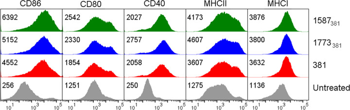
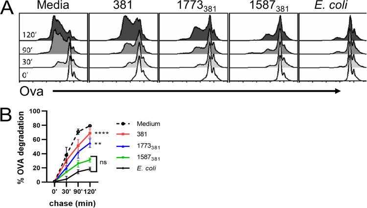
The majority of Gram-negative bacteria elicit a potent immune response via recognition of lipid A expressed on the outer bacterial membrane by the host immune receptor Toll-like receptor 4 (TLR4). However, some Gram-negative bacteria evade detection by TLR4 or alter the outcome of TLR4 signaling by modification of lipid A species. Although the role of lipid A modifications on host innate immunity has been examined in some detail, it is currently unclear how lipid A remodeling influences host adaptive immunity. One prototypic Gram-negative bacterium that modifies its lipid A structure is Porphyromonas gingivalis, an anaerobic pathobiont that colonizes the human periodontium and induces chronic low-grade inflammation that is associated with periodontal disease as well as a number of systemic inflammatory disorders. P. gingivalis produces dephosphorylated and deacylated lipid A structures displaying altered activities at TLR4. Here, we explored the functional role of P. gingivalis lipid A modifications on TLR4-dependent innate and adaptive immune responses in mouse bone marrow-derived dendritic cells (BMDCs). We discovered that lipid A 4'-phosphate removal is required for P. gingivalis to evade BMDC-dependent proinflammatory cytokine responses and markedly limits the bacterium's capacity to induce beta interferon (IFN-β) production. In addition, lipid A 4'-phosphatase activity prevents canonical bacterium-induced delay in antigen degradation, which leads to inefficient antigen cross-presentation and a failure to cross-prime CD8 T cells specific for a P. gingivalis-associated antigen. We propose that lipid A modifications produced by this bacterium alter host TLR4-dependent adaptive immunity to establish chronic infections associated with a number of systemic inflammatory disorders.

Keywords: Porphyromonas gingivalis; TLR4; antigen cross-presentation; bacterial pathogenesis; dendritic cells; inflammation; lipid A.


Figures

FIG 1
FIG 1
Induction of cytokine secretion by P. gingivalis strains. BMDCs differentiated from bone marrow of C57BL/6 or TLR4−/− mice were untreated (medium) or treated for 18 h with P. gingivalis 381 (red), 1773381 (blue), or 1587381 (green) at an MOI of 10, and supernatants were collected and analyzed by ELISA for (A) IL-6, (B) TNF-α, or (C) IL-12p70. Shown are averages from biological triplicates of one experiment (± standard deviation), representative of two independent experiments. Square brackets indicate statistical comparison of P. gingivalis 1587381-treated versus 381- or 1773381-treated C57BL/6 BMDCs. Straight horizontal lines indicate statistical comparison for each treatment of TLR4−/− BMDCs to the correspondingly treated C57BL/6 BMDCs. Statistical analysis was by two-way ANOVA with a Tukey’s multiple-comparison post hoc test. ****, P < 0.0001.
FIG 2
FIG 2
Induction of IFN-β secretion and TLR4 internalization by P. gingivalis strains. BMDCs differentiated from bone marrow of C57/BL6 or TLR4−/− mice were left untreated or were treated for 18 h with P. gingivalis 381 (red), 1773381 (blue), or 1587381 (green) or with heat-killed E. coli cells (black) at the indicated MOI. Supernatants were then collected and analyzed for IFN-β by ELISA. Shown are averages from biological duplicates of one experiment ± standard deviation, representative of four independent experiments. Statistical comparisons are between 1587381 and both 381 and 1773381. Analysis was by two-way ANOVA with a Tukey’s multiple-comparison post hoc test. ****, P < 0.00001.
FIG 3
FIG 3
Upregulation of costimulatory molecules is unaffected by expression of P. gingivalis variant lipid A structures. Surface expression of CD86, CD80, CD40, MHC-II, and MHC-I was assessed by flow cytometry in untreated BMDCs (gray) or after treatment with P. gingivalis 381 (red), 1773381 (blue), or 1587381 (green) for 18 h at an MOI of 10. The numbers in each panel are the geometric mean fluorescent intensity. Shown are results from one experiment representative of three independent experiments.
FIG 4
FIG 4
Effect of lipid A variation on antigen cross-presentation. BMDCs derived from bone marrow of C57BL/6 or TLR4−/− mice were untreated (gray) or treated for 4.5 h with OVA-coated, heat-killed P. gingivalis cells of strain 381 (red), 1773381 (blue), or 1587381 (green) at MOI of 10, 25, or 50 and then cocultured with OT-I T cells. CD69 expression on OT-I T cells was assessed by flow cytometry following an 18-h incubation. As a positive control, unstimulated BMDCs were pulsed with 0.01 ng/ml SIINFEKL peptide before incubation with OT-I cells (black). Data are averages from biological duplicates from one experiment (± standard deviation) representative of three independent experiments. Square brackets indicate statistical comparison of P. gingivalis 1587381-treated versus 381- or 1773381-treated C57BL/6 BMDCs. Straight lines indicate statistical comparison for each treatment of TLR4−/− BMDCs to the correspondingly treated C57BL/6 BMDCs. Statistical analysis was by two-way ANOVA with a Tukey’s multiple-comparison post hoc test. ****, P < 0.0001.
FIG 5
FIG 5
Variation of lipid A structure does not affect uptake of P. gingivalis by BMDCs. C57BL/6 BMDCs were treated with FITC-labeled P. gingivalis strains (MOI of 10) for 30, 60, or 90 min, and bacterial uptake was assessed by flow cytometry. Trypan blue was added to the samples before acquisition to quench extracellular fluorescence due to any membrane-bound bacteria. Shown is one experiment representative of two independent experiments.
FIG 6
FIG 6
Effect of lipid A variation on preservation of internalized antigen. C57BL/6 BMDCs were left untreated (media) or treated with P. gingivalis strains or heat-killed E. coli cells at an MOI of 10 for 16 to 18 h. Bacteria were removed, and cells were pulsed for 10 min with latex beads covalently coupled with OVA. After different periods of time, cells were lysed, and the amount of OVA remaining on beads was assessed by flow cytometry after staining beads for OVA. (A) Histograms showing OVA staining on gated beads for one experiment representative of two independent experiments. (B) Averaged results from two experiments (± standard deviation). The percentage of degradation is 100 − the percentage of undegraded OVA at each time point relative to the percentage undegraded at time zero. Statistical comparisons are for 381 and 1773381 versus 1587381 at 120 min using a two-way ANOVA and Tukey’s multiple-comparison post hoc test. **, P < 0.001; ****, P < 0.0001. There was no statistically significant difference (ns) between 1587381 and E. coli at any time point.
FIG 7
FIG 7
Effect of lipid A variation on cross-priming of OT-I T cells. BMDCs obtained from C57/BL6 or TLR4−/− mice were untreated (gray) or treated for 4.5 h with OVA-coated heat-killed P. gingivalis 381 (red), 1773381 (blue), or 1587381 (green) at MOI of 10, 25, or 50 and then cocultured with OT-I T cells. As a positive control, BMDCs were pulsed with SIINFEKL peptide before addition of T cells (black). Supernatants were collected after an 18-h incubation and analyzed by ELISA for IL-2 (A) and IFN-γ (B). Shown are averages of biological duplicates from one experiment (± standard deviation) representative of three independent experiments. Statistical comparisons are between 1587381 and both 381 and 1773381 using a two-way ANOVA and Tukey’s multiple-comparison post hoc test. # signifies not detected because the IFN-γ concentration was below the level of detection. ****, P < 0.0001.

Similar articles

Cited by

References

    1. Ayres JS. 2016. Cooperative microbial tolerance behaviors in host-microbiota mutualism. Cell 165:1323–1331. doi:10.1016/j.cell.2016.05.049. - DOI - PMC - PubMed
    1. Sansonetti PJ, Di Santo JP. 2007. Debugging how bacteria manipulate the immune response. Immunity 26:149–161. doi:10.1016/j.immuni.2007.02.004. - DOI - PubMed
    1. Simpson BW, Trent MS. 2019. Pushing the envelope: LPS modifications and their consequences. Nat Rev Microbiol 17:403–416. doi:10.1038/s41579-019-0201-x. - DOI - PMC - PubMed
    1. Needham BD, Trent MS. 2013. Fortifying the barrier: the impact of lipid A remodelling on bacterial pathogenesis. Nat Rev Microbiol 11:467–481. doi:10.1038/nrmicro3047. - DOI - PMC - PubMed
    1. Raetz CR, Whitfield C. 2002. Lipopolysaccharide endotoxins. Annu Rev Biochem 71:635–700. doi:10.1146/annurev.biochem.71.110601.135414. - DOI - PMC - PubMed

Publication types

MeSH terms

Substances

LinkOut - more resources