. 2021 Feb 16;89(3):e00309-20.
doi: 10.1128/IAI.00309-20. Print 2021 Feb 16.

STAT3 Promotes Schistosome-Induced Liver Injury by Inflammation, Oxidative Stress, Proliferation, and Apoptosis Signal Pathway

Affiliations

STAT3 Promotes Schistosome-Induced Liver Injury by Inflammation, Oxidative Stress, Proliferation, and Apoptosis Signal Pathway

Jie Zhao et al. Infect Immun. .

Abstract

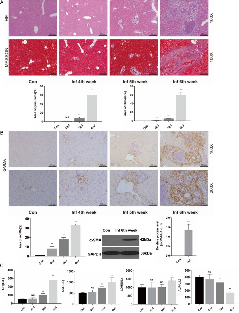
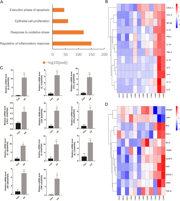
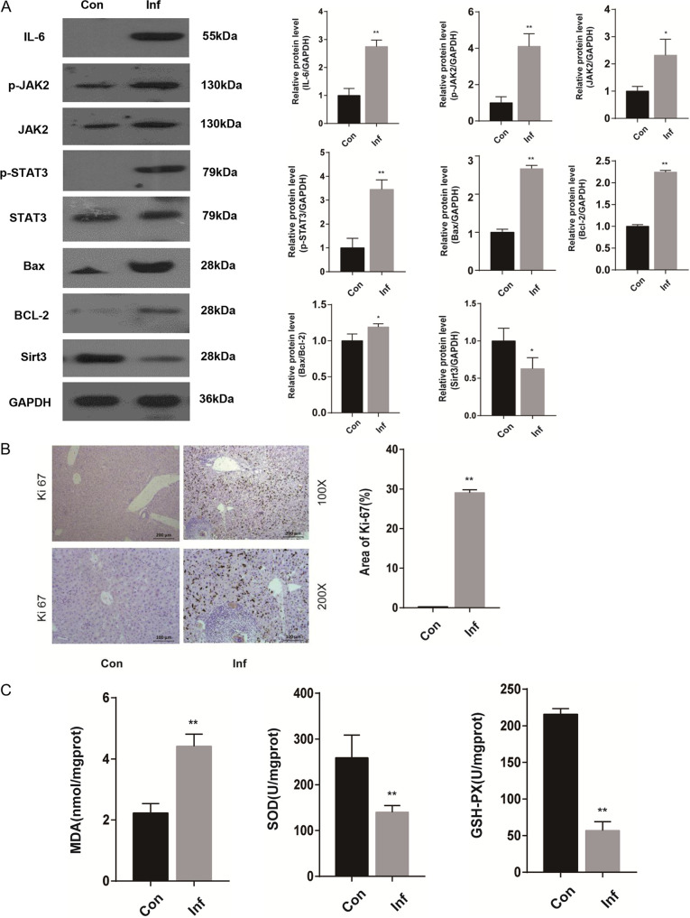
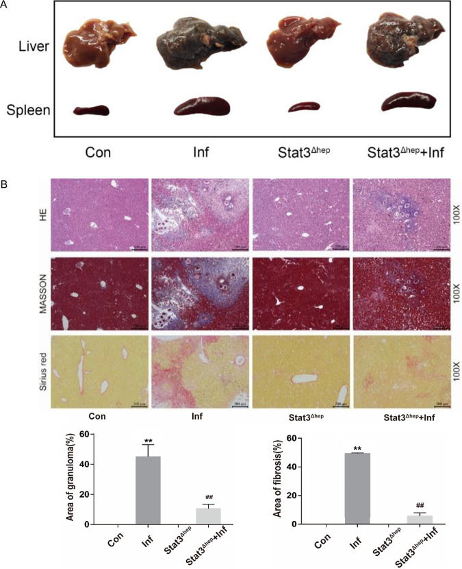
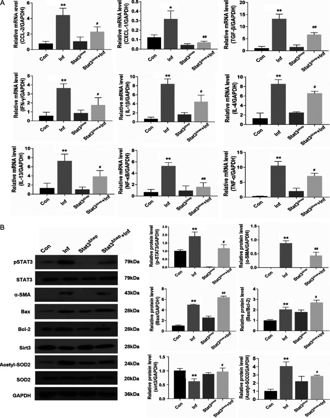
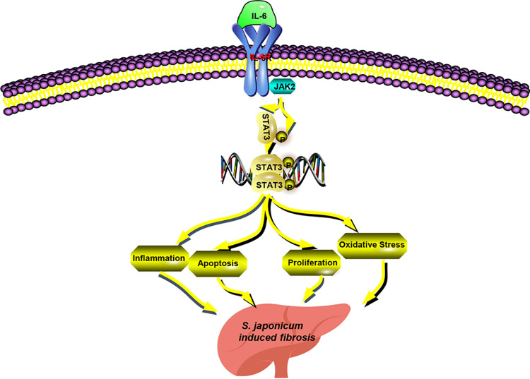
Schistosomiasis is a parasitic helminth disease that can cause organ lesions leading to health damage. During a schistosome infection, schistosome eggs can flow into the liver along the portal vein. Numerous inflammatory cells gather around the eggs, causing granulomas and fibrosis in the liver. In this process, many molecules are involved in the initiation and regulation of the fibrous scar formation. However, the precise molecular mechanisms responsible for the progression of granuloma formation and fibrosis initiation caused by schistosome infection have not been extensively studied. In this study, C57BL/6 wild-type mice and Stat3flox/flox Alb-Cre mice were infected with cercariae of Schistosoma japonicum Liver injury, effector molecule levels, and RNA transcriptome resequencing of liver tissue were detected at 4, 5, and 6 weeks postinfection. We investigated the role of STAT3 (signal transducer and activator of transcription 3) in Schistosoma-induced liver injury in mice. After 6 weeks postinfection, there was obvious liver fibrosis. A sustained pathological process (inflammation, oxidative stress, proliferation, and apoptosis) occurred in S. japonicum-induced liver fibrosis initiation. Meanwhile, we observed activation of the STAT3 pathway in hepatic injury during S. japonicum infection by RNA transcriptome resequencing. Liver deficiency of phospho-STAT3 alleviated infection-induced liver dysfunction, hepatic granuloma formation, and fibrosis initiation. It also promoted STAT3-dependent apoptosis and reduced liver inflammation, oxidative stress, and proliferation. Our results suggest that STAT3 signal pathway and its mediating inflammation, oxidative stress, proliferation, and apoptosis are involved in S. japonicum-induced liver injury and may be a new potential guideline for the treatment of schistosomiasis.

Keywords: S. japonicum; STAT3; apoptosis; inflammation; liver injury; oxidative stress; proliferation.


Figures

FIG 1
FIG 1
Liver granulomas and fibrosis are evident at 6 weeks after S. japonicum infection. Wild-type mice were percutaneously treated with or without 30 ± 2 S. japonicum cercaria. At 4, 5, and 6 weeks postinfection, liver tissue and serum were collected for analysis. (A) H&E staining andMasson’s trichrome staining of liver sections. H&E staining shows the granuloma, and Masson’s trichrome staining shows collagen content and distribution. The granulomatous and fibrotic area as a percentage of total area was measured by computer-assisted morphometric analysis (n = 6). Magnification, ×100. Bar, 200 μm. (B) Representative immunohistochemistry images of α-SMA in liver tissue. Magnification, ×100 (top) and ×200 (bottom). Bars, 200 μm (top) and 100 μm (bottom). Analytical results show the percentage of α-SMA-positive staining in the liver. Western blot analysis of protein levels of α-SMA in liver and quantification (n = 5) are also shown. (C) Serum levels of ALT, AST, LDH, and ALP were measured by use of biochemical analyzer (n = 4). Data are means and SEM for 4 to 6 mice/group. Con, control group; Inf, infected group. * and **, P < 0.05 and P < 0.01 compared to control values. NS, no statistical significance.
FIG 2
FIG 2
Identification of STAT3 involved in regulating host hepatic pathological lesions during S. japonicum infection. Wild-type mice were percutaneously treated with or without 30 ± 2 S. japonicum cercariae. At 4, 5, and 6 weeks postinfection, liver tissue was collected for analysis. (A) GO analysis showing that S. japonicum infection induced the expression of genes involved in several biologic processes (inflammation, oxidative stress, proliferation, and apoptosis) (n = 3). (B) Heat map of the microarray results showing significant upregulation of the inflammatory genes in mice of S. japonicum infection after 4, 5, and 6 weeks compared with control mice. Red, upregulated; blue, downregulated; white, no change. Results are representative of 2 independent experiments (n = 3). (C) Expression of IL-6, IL-11, CCL-2, CXCL-1, TGF-β, IFN-γ, IL-1β, IL-4, IL-13, NF-κB, and TNF-α mRNAs in liver tissue after infection of S. japonicum (n = 4). (D) Heat map of the microarray results showing significant upregulation of the upstream and downstream related genes in mice of S. japonicum infection after 4, 5, and 6 weeks compared with control mice (n = 3). Red, upregulated; blue, downregulated; white, no change. Results are representative of 2 independent experiments. Data are means and SEM for 3 or 4 mice/group. Con, control group; Inf, infected group. * and **, P < 0.05 and P < 0.01 compared to control values.
FIG 3
FIG 3
The JAK2/STAT3 signal pathway is activated in the livers of mice with S. japonicum infection after 6 weeks. Wild-type mice were percutaneously treated with or without 30 ± 2 S. japonicum cercariae. At 6 weeks postinfection, liver tissue was collected for analysis. (A) Western blot analysis and quantification of protein levels of IL-6, p-JAK2, JAK2, p-STAT3, STAT3, Bax, Bcl-2, and Sirt3 in liver. Data are meansand SEM (n = 3 to 6 in each group) from three independent experiments. (B) Representative immunohistochemistry images of Ki-67 in liver tissue. Magnifications, ×100 (top) and ×200 (bottom). Bars, 200 μm (top) and 100 μm (bottom) (n = 4). (C) Measurement of MDA, SOD, and GSH-PX in the liver using a colorimetric reaction with 5,5′-dithiobis-2-nitrobenzoic acid (DTNB) (n = 6). Data are means and SEM for 3 to 6 mice/group. Con, control group; Inf, infected group. * and **, P < 0.05 and P < 0.01 compared to control values.
FIG 4
FIG 4
Liver p-STAT3 deficiency attenuates hepatic injury caused by S. japonicum infection. Wild-type and Stat3Δhep mice were percutaneously treated with or without 30 ± 2 S. japonicum cercariae. At 6 weeks postinfection, liver tissue and serum were collected for analysis. (A) Gross appearance of livers and spleens of control, infected, uninfected Stat3Δhep, and infected Stat3Δhep mice. (B) H&E, Masson’s trichrome, and Sirius red staining of liver sections. H&E staining showed the granuloma, and Masson’s trichrome and Sirius red staining showed collagen content and distribution. The granulomatous and fibrotic area (Masson’s trichrome staining) as a percentage of total area was measured by computer-assisted morphometric analysis (n = 6). Magnification, ×100. Bar, 200 μm. (C) Representative immunohistochemistry images of α-SMA in liver tissue. Magnification, ×100 (top) and ×200 (bottom). Bars, 200 μm (top) and 100 μm (bottom). Analytical results of α-SMA positive staining in the liver (n = 5) are also shown. (D) ALT, AST, LDH, and ALP were measured by use of a biochemical analyzer (n = 4). Data are means and SEM for 3 to 6 mice/group. Con, control group; Inf, infected group; Stat3Δhep: p-STAT3-deficient group; Stat3Δhep+Inf: p-STAT3-deficient infected group. * and **, P < 0.05 and P < 0.01 compared to control values; # and ##, P < 0.05 and P < 0.01 compared to infected-group values. NS, no statistical significance.
FIG 5
FIG 5
Liver p-STAT3 deficiency promotes apoptosis and blocks S. japonicum-induced hepatic inflammation, proliferation, and oxidative stress. Wild-type and Stat3Δhep mice were percutaneously treated with or without 30 ± 2 S. japonicum cercariae. At 6 weeks postinfection, liver tissues were analyzed. (A)Expression of CCL-2, CXCL-1, TGF-β, IFN-γ, IL-1β, IL-4, IL-13, NF-κB, and TNF-α mRNAs in liver tissue after infection of S. japonicum (n = 4). (B) Western blot analysis and quantification of protein levels of p-STAT3, STAT3, α-SMA, Bax, Bcl-2, Sirt3, acetyl-SOD2, and SOD2 in liver. Data are means and SEM (n = 3 to 6 in each group) from three independent experiments (n = 3). (C) Representative immunohistochemistry images of Ki-67 in liver tissue. Magnification, ×100 (top) and ×200 (bottom). Bars, 200 μm (top) and 100 μm (bottom) (n = 4). (D) Measurement of MDA, SOD, and GSH-PX in the liver using a colorimetric reaction with DNTB (n = 6). Data are means and SEM for 3 to 6 mice/group. Con, control group; Inf, infected group; Stat3Δhep, uninfected p-STAT3-deficient group; Stat3Δhep+Inf, infected p-STAT3-deficient group. * and **, P < 0.05 and P < 0.01 compared with control values; # and ##, P < 0.05 and P < 0.01 compared with infected-group values; NS, no statistical significance.
FIG 6
FIG 6
Schematic overview of JAK2/STAT3-mediated effects on S. japonicum-induced liver fibrosis. The JAK2/STAT3 signal pathway and its related molecules, such as IL-6, could be activated in the livers of mice with S. japonicum infection. STAT3 constitutes a central node responsible for inflammation, apoptosis, proliferation, and oxidative stress by regulating various downstream targets that ultimately favor promotion of liver fibrosis.

Similar articles

Cited by

References

    1. Colley DG, Bustinduy AL, Secor WE, King CH. 2014. Human schistosomiasis. Lancet 383:2253–2264. doi:10.1016/S0140-6736(13)61949-2. - DOI - PMC - PubMed
    1. Liu X, Zhang YR, Cai C, Ni XQ, Zhu Q, Ren JL, Chen Y, Zhang LS, Xue CD, Zhao J, Qi YF, Yu YR. 2019. Taurine alleviates Schistosoma-induced liver injury by inhibiting the TXNIP/NLRP3 inflammasome signal pathway and pyroptosis. Infect Immun 87:e00732-19. doi:10.1128/IAI.00732-19. - DOI - PMC - PubMed
    1. Zhan TZ, Ma HH, Jiang SQ, Zhong ZR, Wang XL, Li CX, Yu D, Liu L, Xu J, Xia CM. 2019. IL-9 blockage reduces early hepatic granuloma formation and fibrosis during Schistosoma japonicum infection in mice. Immunology 158:296–303. doi:10.1111/imm.13111. - DOI - PMC - PubMed
    1. Barsoum RS, Esmat G, El-Baz T. 2013. Human schistosomiasis: clinical perspective: review. J Adv Res 4:433–444. doi:10.1016/j.jare.2013.01.005. - DOI - PMC - PubMed
    1. He X, Wang Y, Fan XB, Lei NH, Tian YN, Zhang DM, Pan WQ. 2020. A schistosome miRNA promotes host hepatic fibrosis by targeting transforming growth factor beta receptor III. J Hepatol 72:519–527. doi:10.1016/j.jhep.2019.10.029. - DOI - PubMed

Publication types

MeSH terms

Substances

LinkOut - more resources