Observational Study
. 2020 Dec 15;117(50):31674-31684.
doi: 10.1073/pnas.2019786117. Epub 2020 Nov 30.

A wireless, skin-interfaced biosensor for cerebral hemodynamic monitoring in pediatric care

Affiliations
Observational Study

A wireless, skin-interfaced biosensor for cerebral hemodynamic monitoring in pediatric care

Alina Y Rwei et al. Proc Natl Acad Sci U S A. .

Abstract

The standard of clinical care in many pediatric and neonatal neurocritical care units involves continuous monitoring of cerebral hemodynamics using hard-wired devices that physically adhere to the skin and connect to base stations that commonly mount on an adjacent wall or stand. Risks of iatrogenic skin injuries associated with adhesives that bond such systems to the skin and entanglements of the patients and/or the healthcare professionals with the wires can impede clinical procedures and natural movements that are critical to the care, development, and recovery of pediatric patients. This paper presents a wireless, miniaturized, and mechanically soft, flexible device that supports measurements quantitatively comparable to existing clinical standards. The system features a multiphotodiode array and pair of light-emitting diodes for simultaneous monitoring of systemic and cerebral hemodynamics, with ability to measure cerebral oxygenation, heart rate, peripheral oxygenation, and potentially cerebral pulse pressure and vascular tone, through the utilization of multiwavelength reflectance-mode photoplethysmography and functional near-infrared spectroscopy. Monte Carlo optical simulations define the tissue-probing depths for source-detector distances and operating wavelengths of these systems using magnetic resonance images of the head of a representative pediatric patient to define the relevant geometries. Clinical studies on pediatric subjects with and without congenital central hypoventilation syndrome validate the feasibility for using this system in operating hospitals and define its advantages relative to established technologies. This platform has the potential to substantially enhance the quality of pediatric care across a wide range of conditions and use scenarios, not only in advanced hospital settings but also in clinics of lower- and middle-income countries.

Keywords: bioelectronics; cerebral hemodynamics; near-infrared spectroscopy; wearable electronics.


Conflict of interest statement

The authors declare no competing interest.

Figures
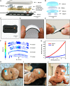
Fig. 1.
Fig. 1.
Design and mechanical characterization of a soft, wireless device for cerebral hemodynamic monitoring on pediatric subjects. (A and B) Schematic illustration and exploded view of (A) a device with (B) a modular, rechargeable coin cell battery. The main body consists of serpentine interconnects between islands of electrical components for wireless operation and optical tissue profiling, with four photodiodes each featuring a distinct source–detector distance. The main body and the modular battery both feature a pair of magnets for mechanical and electrical coupling, all encapsulated within soft, medical-grade silicone. (C) Photograph of the skin-oriented side of the main device unit, with a pair of LEDs and four photodetectors encapsulated between a black silicone optical barrier and coated with transparent PDMS for effective optical coupling. (D and E) Photograph of a device upon mechanical stresses of (D) bending and (E) twisting. (F) Computed strain distribution in the copper layers (first Cu layer is the closest to skin) of the device with a bending radius of 2.5 cm. (G) Mechanical characterization of the device main unit compared with pig skin using a three-point flexural test. (HJ) Photographs of a device with a modular battery mounted on the forehead of a 9-wk-old infant during (H) resting, (I) feeding, and (J) tummy time.
Fig. 2.
Fig. 2.
Optical distribution of NIR light in the cerebral tissue. (A and B) Representative MRI data from a term-born infant and manually defined scalp (coral), skull (light gray), cerebral spinal fluid (blue), gray matter (dark gray), and white matter (gray) in the (A) axial view and (B) sagittal view. (C) Computed optical distribution of NIR light (850 nm) via Monte Carlo simulation (red: light source; green: photodetectors). (D) Relative partial pathlength of light at each source–detector distance within each respective tissue type.
Fig. 3.
Fig. 3.
Operational characteristics of our device for cerebral hemodynamics monitoring. (A) Schematic block diagram for our BLE electronic module. The module contains the LEDs and the driving circuit, PDs, and transimpedance amplifier circuits, a BLE microcontroller with a custom-developed program, and peripheral circuits. (B) GUI for the cerebral oximeter. The GUI supports real-time visualization, storage, and analysis of measurement data and provides a control interface for setting parameters for the BLE module. (C) Flowchart of the data analysis procedure for extraction of cerebral oxygenation (ScO2), peripheral oxygenation (SpO2), HR, and cerebral vascular tone. LPF = low-pass filter, BPF= band-pass filter, R = red, and IR = infrared. PD1, PD2, PD3, and PD4 correspond to source–detector distances of 5, 10, 15, and 20 mm respectively.
Fig. 4.
Fig. 4.
Data collection of systemic and cerebral hemodynamics from healthy young adults. (A) Representative cerebral oxygenation measurements from our device and a commercialized NIRS instrument during end inspiratory breath-hold studies on a 20-y-old African American subject. (B) Bland–Altman analysis of cerebral oxygenation measurements between our device and a commercialized NIRS instrument (n = 3 subjects, three breath-hold events per subject). (C) Representative pulse oxygenation measurements from our device and a standard SpO2 finger probe during end inspiratory breath-hold studies on a 29-y-old subject. (D) Bland–Altman analysis of pulse oxygenation measurements between our device and a commercialized SpO2 finger probe (n = 3 subjects, three breath-hold events per subject). (E) Representative HR analysis from our device and a clinically used finger probe during end inspiratory breath-hold on a 19-y-old subject. (F) Bland–Altman analysis of HR analyses between our device and a medical-grade finger probe during end inspiratory breath-hold exercises (n = 3 subjects, three breath-hold events per subject). (G and H) Representative vascular tone and pulse pressure measurements from our device, reflected from the change of peak amplitude, during (G) end inspiratory breath-hold (eight events) and (H) breath-in (defined as deep inspiration, followed by immediate expiration, five events) exercises on a 16-y-old subject.
Fig. 5.
Fig. 5.
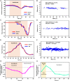
Data collection from pediatric subjects with and without CCHS. (A) Representative NIRS measurements from our device and medical-grade NIRS on pediatric subjects during a HUT challenge on an 8-y-old African American boy with CCHS. (B) Relative changes in cerebral oxygenation of pediatric subjects measured by our device and a commercialized NIRS system during tilt phase and recovery phase relative to a 10-min baseline period (n = 10). Statistical analysis was performed by a paired Student t test. *P < 0.01 when compared with baseline. A P value <0.05 was considered statistically significant. (C) Representative NIRS measurements during a hyperoxia challenge with 100% oxygen on a 4-y-old Caucasian girl with CCHS. (D) Representative NIRS measurements during a hypoxia challenge with 5 breaths of 100% nitrogen gas on a 13-y-old Hispanic girl with CCHS. (E) Photograph of our device on the 2-mo-old infant upon cerebral oxygenation measurements during normal activity. (F) Bland–Altman analysis of our device compared with a medical-grade NIRS device from HUT, hyperoxia (100% O2), hypoxia challenges (100% N2), and hypoxic–hypercapnia (14% O2 and 7% CO2 balanced with nitrogen [N2]) with pediatric subjects aged 4 to 15 y and during normal activity (NA) with pediatric subjects aged 0.2 to 2 y.

References

    1. Carroll M. S., et al. , Residual chemosensitivity to ventilatory challenges in genotyped congenital central hypoventilation syndrome. J Appl Physiol (1985) 116, 439–450 (2014). - PubMed
    1. Vesoulis Z. A., Mathur A. M., Cerebral autoregulation, brain injury, and the transitioning premature infant. Front Pediatr. 5, 64 (2017). - PMC - PubMed
    1. Zaleski K. L., Kussman B. D., Near-infrared spectroscopy in pediatric congenital heart disease. J. Cardiothorac. Vasc. Anesth. 34, 489–500 (2020). - PubMed
    1. Yu Y., et al. , Cerebral near-infrared spectroscopy (NIRS) for perioperative monitoring of brain oxygenation in children and adults. Cochrane Database Syst. Rev. 1, CD010947 (2018). - PMC - PubMed
    1. Serena S. B. S., et al. , Young age as a risk factor for impaired cerebral autoregulation after moderate to severe pediatric traumatic brain injury. Anesthesiology J. Am. Soc. Anesthesiologists 108, 588–595 (2008). - PubMed

Publication types

MeSH terms

LinkOut - more resources