. 2020 Dec 15;117(50):31824-31831.
doi: 10.1073/pnas.2013904117. Epub 2020 Nov 30.

Viewing rare conformations of the β2 adrenergic receptor with pressure-resolved DEER spectroscopy

Affiliations

Viewing rare conformations of the β2 adrenergic receptor with pressure-resolved DEER spectroscopy

Michael T Lerch et al. Proc Natl Acad Sci U S A. .

Abstract

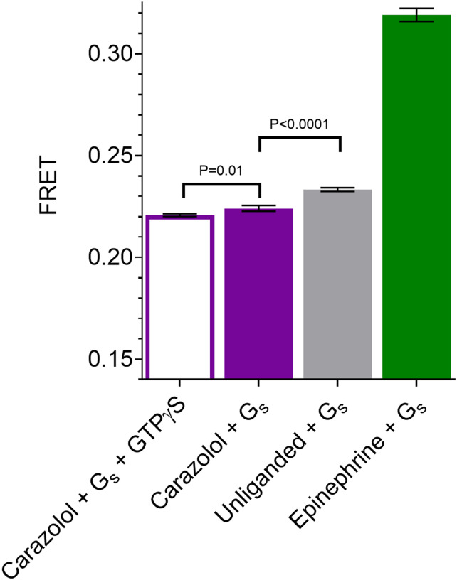
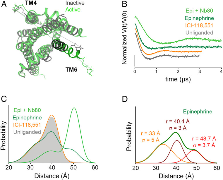
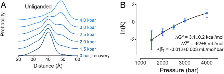
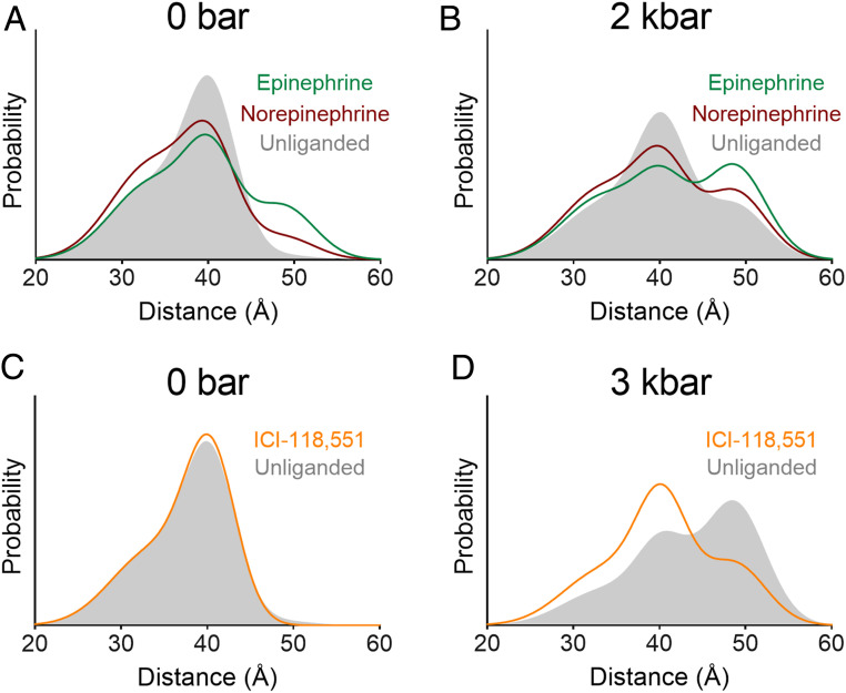
The β2 adrenergic receptor (β2AR) is an archetypal G protein coupled receptor (GPCR). One structural signature of GPCR activation is a large-scale movement (ca. 6 to 14 Å) of transmembrane helix 6 (TM6) to a conformation which binds and activates a cognate G protein. The β2AR exhibits a low level of agonist-independent G protein activation. The structural origin of this basal activity and its suppression by inverse agonists is unknown but could involve a unique receptor conformation that promotes G protein activation. Alternatively, a conformational selection model proposes that a minor population of the canonical active receptor conformation exists in equilibrium with inactive forms, thus giving rise to basal activity of the ligand-free receptor. Previous spin-labeling and fluorescence resonance energy transfer experiments designed to monitor the positional distribution of TM6 did not detect the presence of the active conformation of ligand-free β2AR. Here we employ spin-labeling and pressure-resolved double electron-electron resonance spectroscopy to reveal the presence of a minor population of unliganded receptor, with the signature outward TM6 displacement, in equilibrium with inactive conformations. Binding of inverse agonists suppresses this population. These results provide direct structural evidence in favor of a conformational selection model for basal activity in β2AR and provide a mechanism for inverse agonism. In addition, they emphasize 1) the importance of minor populations in GPCR catalytic function; 2) the use of spin-labeling and variable-pressure electron paramagnetic resonance to reveal them in a membrane protein; and 3) the quantitative evaluation of their thermodynamic properties relative to the inactive forms, including free energy, partial molar volume, and compressibility.

Keywords: basal activity; conformational selection; double electron–electron resonance; high pressure; β2 adrenergic receptor.


Conflict of interest statement

Competing interest statement: B.K.K. is a cofounder of and consultant for ConfometRx, Inc.

Figures

Fig. 1.
Fig. 1.
FRET measurement of basal activity of the β2AR. β2AR-Gs complex formation was monitored under buffer conditions similar to DEER sample conditions, either without ligand or in the presence of saturating concentrations of epinephrine or carazolol. FRET experiments on β2AR labeled with Cy3B (donor fluorophore) and Gs labeled with Cy5 (acceptor fluorophore) reveal the FRET intensity due to β2AR-Gs complex formation in the presence or absence of an excess of the nonhydrolyzable GTP analog GTPγS (Materials and Methods). Error bars denote SD, four replicates. Statistical significance is tested with Welch’s t test.
Fig. 2.
Fig. 2.
Monitoring the β2AR TM6 conformational equilibrium with DEER. (A) Crystal structures of inactive (gray; Protein Data Bank [PDB] ID 2RH1) and active (green; PDB ID 3SN6) β2AR with a PROXYL side chain shown at labeled sites in TM4 (148) and TM6 (266). (B) Background-corrected DEFs and fits for the unliganded receptor as well as receptor bound to an inverse agonist (ICI-118,551), agonist (epinephrine), and agonist + G protein mimic (Nb80) at atmospheric pressure. The data are displayed as dots and the fits as solid lines. (C) Distance distributions obtained from fits of the data to a Gaussian model. (D) Individual Gaussian components of the distance distribution for the receptor bound to epinephrine. All pressure-dependent and ligand-bound DEER distance distributions are composed of these three components with varying amplitude.
Fig. 3.
Fig. 3.
Pressure increases the population of the active conformation in unliganded β2AR. (A) Distance distributions of unliganded receptor as a function of pressure, as well as the recovery distance distribution (dotted black line) collected for the 4-kbar sample after allowing it to reequilibrate at atmospheric pressure. (B) Plot of ln(K) vs. pressure and fit to a two-state model to yield the difference in free energy (ΔGo), partial molar volume changes (ΔV¯o), and compressibility (Δβ¯T) for the inactive-to-active transition. DEFs for these samples are shown in SI Appendix, Fig. S3.
Fig. 4.
Fig. 4.
β2AR pressure response is ligand-dependent. Distance distributions for the unliganded receptor overlaid with receptor bound to endogenous agonists at (A) atmospheric pressure and (B) 2 kbar, as well as inverse agonist (ICI-118,551) at (C) atmospheric pressure and (D) 3 kbar. Ligands are present at saturating concentrations in each sample. DEFs for these samples are shown in SI Appendix, Fig. S5.

References

    1. Cherezov V., et al. , High-resolution crystal structure of an engineered human β2-adrenergic G protein-coupled receptor. Science 318, 1258–1265 (2007). - PMC - PubMed
    1. Altenbach C., Kusnetzow A. K., Ernst O. P., Hofmann K. P., Hubbell W. L., High-resolution distance mapping in rhodopsin reveals the pattern of helix movement due to activation. Proc. Natl. Acad. Sci. U.S.A. 105, 7439–7444 (2008). - PMC - PubMed
    1. Rasmussen S. G. F., et al. , Crystal structure of the β2 adrenergic receptor-Gs protein complex. Nature 477, 549–555 (2011). - PMC - PubMed
    1. Leurs R., Smit M. J., Alewijnse A. E., Timmerman H., Agonist-independent regulation of constitutively active G-protein-coupled receptors. Trends Biochem. Sci. 23, 418–422 (1998). - PubMed
    1. Zhou Y.-Y., et al. , Spontaneous activation of β(2)- but not β(1)-adrenoceptors expressed in cardiac myocytes from β(1)β(2) double knockout mice. Mol. Pharmacol. 58, 887–894 (2000). - PubMed

Publication types

MeSH terms

Substances