. 2020 Dec 15;117(50):32056-32065.
doi: 10.1073/pnas.2005877117. Epub 2020 Nov 30.

Mitochondrial Nuclear Retrograde Regulator 1 (MNRR1) rescues the cellular phenotype of MELAS by inducing homeostatic mechanisms

Affiliations

Mitochondrial Nuclear Retrograde Regulator 1 (MNRR1) rescues the cellular phenotype of MELAS by inducing homeostatic mechanisms

Siddhesh Aras et al. Proc Natl Acad Sci U S A. .

Erratum in

Abstract

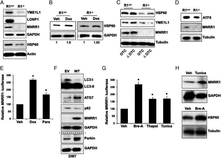
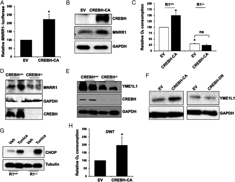
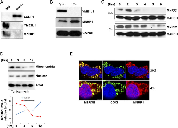
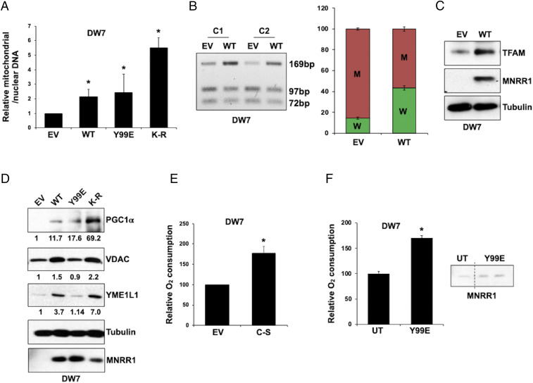
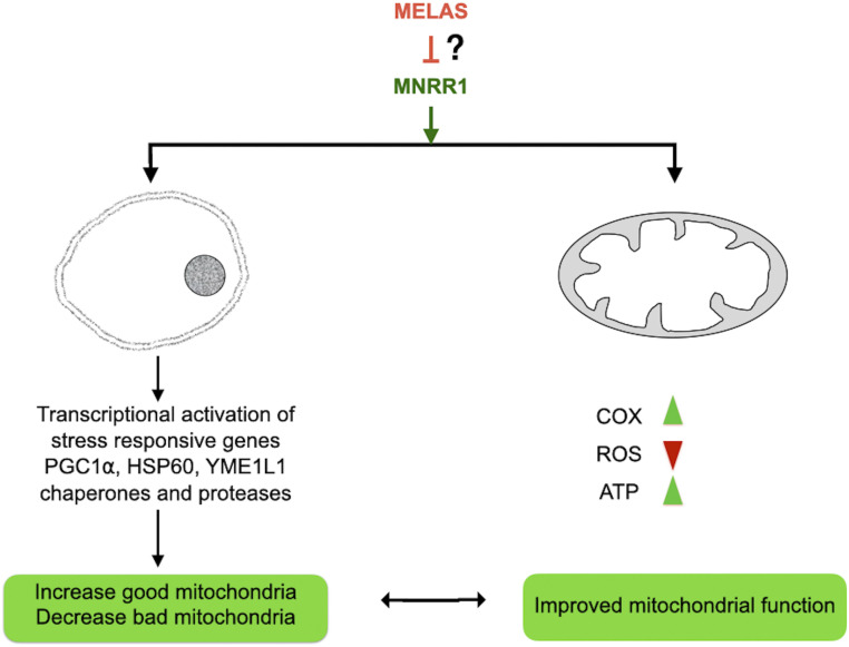
MNRR1 (CHCHD2) is a bi-organellar regulator of mitochondrial function that directly activates cytochrome c oxidase in the mitochondria and functions in the nucleus as a transcriptional activator for hundreds of genes. Since MNRR1 depletion contains features of a mitochondrial disease phenotype, we evaluated the effects of forced expression of MNRR1 on the mitochondrial disease MELAS (mitochondrial encephalomyopathy, lactic acidosis and stroke-like episodes) syndrome. MELAS is a multisystem encephalomyopathy disorder that can result from a heteroplasmic mutation in the mitochondrial DNA (mtDNA; m.3243A > G) at heteroplasmy levels of ∼50 to 90%. Since cybrid cell lines with 73% m.3243A > G heteroplasmy (DW7) display a significant reduction in MNRR1 levels compared to the wild type (0% heteroplasmy) (CL9), we evaluated the effects of MNRR1 levels on mitochondrial functioning. Overexpression of MNRR1 in DW7 cells induces the mitochondrial unfolded protein response (UPRmt), autophagy, and mitochondrial biogenesis, thereby rescuing the mitochondrial phenotype. It does so primarily as a transcription activator, revealing this function to be a potential therapeutic target. The role of MNRR1 in stimulating UPRmt, which is blunted in MELAS cells, was surprising and further investigation uncovered that under conditions of stress the import of MNRR1 into the mitochondria was blocked, allowing the protein to accumulate in the nucleus to enhance its transcription function. In the mammalian system, ATF5, has been identified as a mediator of UPRmt MNRR1 knockout cells display an ∼40% reduction in the protein levels of ATF5, suggesting that MNRR1 plays an important role upstream of this known mediator of UPRmt.

Keywords: CHCHD2; cytochrome c oxidase; mitochondria; transcription; unfolded protein response.


Conflict of interest statement

Competing interest statement: Company associations for D.C.W. not related to the manuscript include MitoCURia, Mitrios, and Panos.

Figures

Fig. 1.
Fig. 1.
MNRR1 expression in the deficient MELAS cells rescues mitochondrial function. (A) MNRR1 and mitochondrial ETC complex levels in CL9 (100% WT mtDNA) and DW7 (73% mutant mtDNA) MELAS cybrid cells. GAPDH served as a loading control. MNRR1 transcript levels in CL9 and DW7 cells normalized to GAPDH are also shown. (B) Intact cellular oxygen consumption in the DW7 MELAS cell lines expressing an EV, WT MNRR1, or the mitochondrially active (Y99E) and transcriptionally active (K-R) mutants of MNRR1. Data are represented as oxygen consumption relative to CL9 cells expressing the EV set to 100%. (C) ATP levels in DW7 cells expressing either an EV or WT MNRR1. (D) Total cellular ROS levels in DW7 cells expressing an EV, WT MNRR1, or the transcriptionally active mutant (K-R). (E) Immunofluorescence microscopy in DW7 cells expressing an EV or WT MNRR1, each along with Mito-mCherry and stained with MitoTracker Green. The Pearson correlation coefficient for yellow overlap intensity is also shown. (F) DW7 MELAS cells are defective for their ability to induce UPRmt. CL9 (100% WT mtDNA) and DW7 (73% mutant mtDNA) MELAS cells were treated with either vehicle (Veh) or doxycycline (Dox; 50 μg/mL), and YME1L1 levels were determined. Relative MNRR1 band density is shown beneath. GAPDH served as a loading control. (G) MNRR1-expressing DW7 cells show an increase in the YME1L1 protein levels compared with cells expressing an EV. GAPDH served as a loading control.
Fig. 2.
Fig. 2.
Cellular stress activates MNRR1, which is required for optimal induction of UPRmt. (A) Protein levels of key UPRmt markers in MNRR1-KO HEK 293 cells (R1−/−) compared with control cells (R1+/+). GAPDH and actin served as loading controls. (B) Induction of UPRmt is deficient in MNRR1-KO cells (R1−/−) on doxycycline treatment (50 μg/mL) for 48 h. The ratio of HSP60/GAPDH intensity is shown beneath. (C) Induction of UPRmt is deficient in MNRR1-KO cells (R1−/−) when the deletion mutant of ornithine transcarbamylase (ΔOTC), but not of WT (OTC), is expressed. HSP60 and YME1L1 are probed as markers of UPRmt with tubulin as a loading control. (D) ATF5 levels in MNRR1-KO HEK 293 cells (R1−/−) compared with control cells (R1+/+). Tubulin served as a loading control. (E) Dual luciferase assay in HEK 293 cells expressing the MNRR1-luciferase reporter treated with UPRmt inducers doxycycline (50 μg/mL) and paraquat (10 μM) for 48 h. Data are presented relative to cells treated with vehicle. (F) Levels of autophagy markers in DW7 MELAS cells expressing either an EV or WT MNRR1. GAPDH served as a loading control. MNRR1 expression was detected by a Flag antibody. (G) Dual luciferase assay in cells expressing the MNRR1-luciferase reporter treated with ER stress inducer brefeldin A (Bre-A) (100 μg/mL), thapsigargin (1 μM), or tunicamycin (5 μg/mL). Data are represented relative to cells treated with vehicle. (H) MNRR1 protein levels in cells treated with 5 μg/mL tunicamycin (Tunica), with GAPDH as a loading control, and UPRmt marker (HSP60) levels in cells treated with Bre-A (100 μg/mL), a chemical inducer of ER stress, with tubulin as a loading control.
Fig. 3.
Fig. 3.
CREBH, an ER stress-responsive transcription factor, activates MNRR1. (A) Transcription measured by a dual luciferase assay in HEK 293 cells coexpressing the MNRR1-luciferase reporter with either an EV or CREBH-CA. Data are presented as luciferase levels relative to EV-expressing cells. (B) MNRR1 protein levels in HEK 293 cells expressing either an EV or CREBH-CA. GAPDH served as a loading control. (C) Cellular oxygen consumption in intact MNRR1-KO HEK 293 cells (R1−/−) compared with control cells (R1+/+) expressing either an EV or CREBH-CA. Oxygen consumption is shown relative to R1+/+ cells expressing the EV. (D) MNRR1 protein levels in CREBH WT (CREBH+/+) and KO (CREBH−/−) mouse liver tissues (n = 2). (E) UPRmt marker YME1L1 protein levels in CREBH WT (CREBH+/+) and KO (CREBH−/−) mouse liver tissues (n = 2). (F) YME1L1 protein levels in HEK 293 cells expressing either CREBH-CA or CREBH-DN compared with cells expressing an EV. GAPDH served as a loading control. (G) ER stress marker CHOP protein levels in MNRR1-KO HEK 293 cells (R1−/−) compared with control cells (R1+/+) treated with either vehicle or 5 μg/mL tunicamycin (Tunica). (H) Cellular oxygen consumption in intact DW7 cells expressing either an EV or CREBH-CA. Oxygen consumption is shown relative to cells expressing the EV.
Fig. 4.
Fig. 4.
Mitochondrial import of MNRR1 is inhibited under stress. (A) Submitochondrial localization of MNRR1 along with UPRmt markers YME1L1 and LonP1. (B) MNRR1 levels in YME1L1-KO (Y−/−) MEFs compared with WT (Y+/+) cells. GAPDH served as a loading control. (C) Cycloheximide (CHX) chase experiment in WT (Y+/+) and YME1L1-KO (Y−/−) MEFs for MNRR1 levels. GAPDH served as a loading control. Cells were treated with 100 μg/mL CHX for the indicated times. (D) YME1L1−/− HEK 293 cells were treated with tunicamycin (5 μg/mL) for the indicated times. In addition to total cell lysate, treated cells were fractionated into mitochondrial and nuclear fractions, and MNRR1 levels were determined. The graph depicts the ratio of organellar fraction to total cellular MNRR1 intensity at the indicated time points. (E) Immunofluorescent confocal microscopy for MNRR1 localization in MEFs maintained for 8 h at either 20% or 4% oxygen. COX1 (green) was used as a mitochondrial marker, and DAPI (blue) was used to stain the nucleus. MNRR1 staining is in red.
Fig. 5.
Fig. 5.
Effects of MNRR1 expression in DW7 MELAS cells. (A) Ratio of mitochondrial DNA to nuclear DNA in DW7 MELAS cells expressing an EV, WT MNRR1, mitochondrially active (Y99E), or transcriptionally active (K-R) mutants of MNRR1. The ratio is relative to the EV value set to 1. (B) HaeIII restriction enzyme digestion of a PCR-amplified fragment of mtDNA harboring the 3234 mutation from DW7 cells stably expressing either an EV or WT MNRR1. Quantitation is depicted in the bar graph. W indicates WT mtDNA; M, mutant mtDNA. Two independent clones for both EV and WT (clones 1 and 2) were tested for heteroplasmy level; the bar graph shows the average of the two values. For EV, the values for clone 1 were 14.4% WT and 85.6% mutant; for WT, these values were 43.6% WT and 56.4% mutant. (C) WT MNRR1 expression increases TFAM levels in DW7 MELAS cells compared with those expressing the EV. (D) Levels of PGC1α (mitochondrial biogenesis), VDAC (mitochondrial amount), and YME1L1 (UPRmt) markers in DW7 cells expressing the EV, WT MNRR1, and the mitochondrially active (Y99E) or transcriptionally active (K-R) mutants of MNRR1. Tubulin served as a loading control. Band densities relative to the EV column are shown. MNRR1 expression was detected by a Flag antibody. (E) Nuclear- localized mutant of MNRR1 (C-S) increases mitochondrial oxygen consumption in DW7 MELAS cells compared with the EV. (F) Peptide therapy rescues function in DW7 MELAS cells. (Left) The recombinant mitochondrially active Y99E mutant protein was transfected into DW7 MELAS cells and was able to increase mitochondrial oxygen consumption. (Right) Increase in MNRR1 protein in Y99E transfected cells versus nontransfected cells (UT).
Fig. 6.
Fig. 6.
Model for the mechanism of MNRR1-mediated rescue of mitochondrial function in MELAS. Via its mitochondrial function, MNRR1 increases organellar respiratory activity, and via its nuclear function, it promotes mitochondrial biogenesis and induction of quality control physiological processes, such as autophagy, to restore homeostasis.

Similar articles

Cited by

References

    1. Aras S., et al. , Abl2 kinase phosphorylates bi-organellar regulator MNRR1 in mitochondria, stimulating respiration. Biochim. Biophys. Acta Mol. Cell Res. 1864, 440–448 (2017). - PubMed
    1. Aras S., et al. , MNRR1 (formerly CHCHD2) is a bi-organellar regulator of mitochondrial metabolism. Mitochondrion 20, 43–51 (2015). - PubMed
    1. Aras S., et al. , Oxygen-dependent expression of cytochrome c oxidase subunit 4-2 gene expression is mediated by transcription factors RBPJ, CXXC5, and CHCHD2. Nucleic Acids Res. 41, 2255–2266 (2013). - PMC - PubMed
    1. Liu Y., et al. , CHCHD2 inhibits apoptosis by interacting with Bcl-xL to regulate Bax activation. Cell Death Differ. 22, 1035–1046 (2015). - PMC - PubMed
    1. Kirby D. M., et al. , Mutations of the mitochondrial ND1 gene as a cause of MELAS. J. Med. Genet. 41, 784–789 (2004). - PMC - PubMed

Publication types

MeSH terms

LinkOut - more resources