. 2020 Dec 15;117(50):32155-32164.
doi: 10.1073/pnas.2008456117. Epub 2020 Nov 30.

Targeting presynaptic H3 heteroreceptor in nucleus accumbens to improve anxiety and obsessive-compulsive-like behaviors

Affiliations

Targeting presynaptic H3 heteroreceptor in nucleus accumbens to improve anxiety and obsessive-compulsive-like behaviors

Xiao-Yang Zhang et al. Proc Natl Acad Sci U S A. .

Abstract

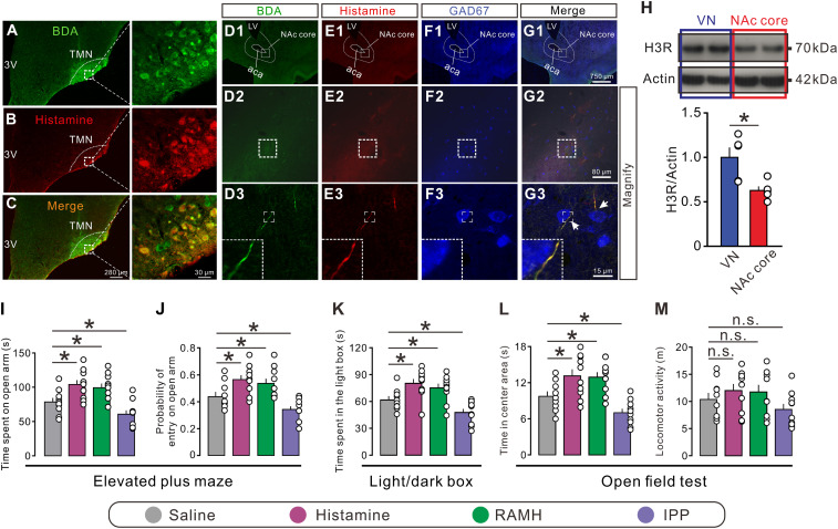
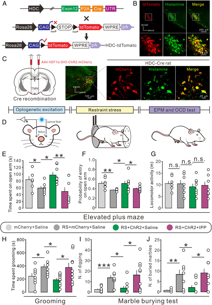
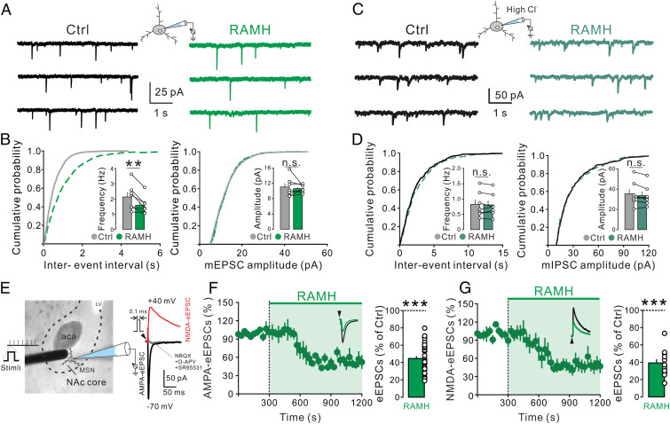
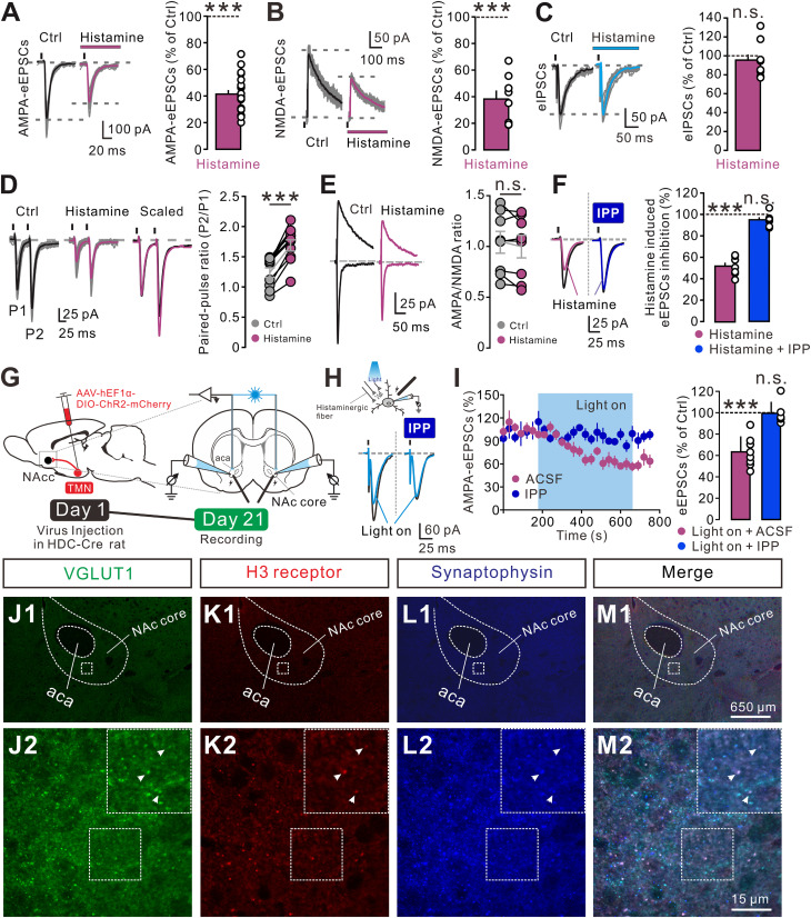
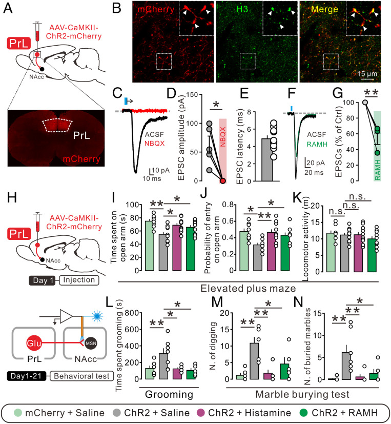
Anxiety commonly co-occurs with obsessive-compulsive disorder (OCD). Both of them are closely related to stress. However, the shared neurobiological substrates and therapeutic targets remain unclear. Here we report an amelioration of both anxiety and OCD via the histamine presynaptic H3 heteroreceptor on glutamatergic afferent terminals from the prelimbic prefrontal cortex (PrL) to the nucleus accumbens (NAc) core, a vital node in the limbic loop. The NAc core receives direct hypothalamic histaminergic projections, and optogenetic activation of hypothalamic NAc core histaminergic afferents selectively suppresses glutamatergic rather than GABAergic synaptic transmission in the NAc core via the H3 receptor and thus produces an anxiolytic effect and improves anxiety- and obsessive-compulsive-like behaviors induced by restraint stress. Although the H3 receptor is expressed in glutamatergic afferent terminals from the PrL, basolateral amygdala (BLA), and ventral hippocampus (vHipp), rather than the thalamus, only the PrL- and not BLA- and vHipp-NAc core glutamatergic pathways among the glutamatergic afferent inputs to the NAc core is responsible for co-occurrence of anxiety- and obsessive-compulsive-like behaviors. Furthermore, activation of the H3 receptor ameliorates anxiety and obsessive-compulsive-like behaviors induced by optogenetic excitation of the PrL-NAc glutamatergic afferents. These results demonstrate a common mechanism regulating anxiety- and obsessive-compulsive-like behaviors and provide insight into the clinical treatment strategy for OCD with comorbid anxiety by targeting the histamine H3 receptor in the NAc core.

Keywords: OCD; anxiety; histamine H3 receptor; nucleus accumbens; prelimbic prefrontal cortex.


Conflict of interest statement

The authors declare no competing interest.

Figures

Fig. 1.
Fig. 1.
Histaminergic afferents in the NAc core and an involvement of the H3 receptor in anxiety-related behaviors. (AC) Immunostaining micrographs showing the identification of histaminergic neurons with injections of BDA into the TMN. (D1 to G3) Triple immunostaining shows that the anterogradely labeled BDA fibers (green) in the NAc core contain immunoreactivity for histamine (red). Note that these histaminergic fibers pass around GAD67-labeled GABAergic neurons (blue) in the NAc core. LV, lateral ventricle; 3V, third ventricle; aca, anterior commissure, anterior part. (H) Western blot analysis indicates that the histamine H3 receptor is expressed in the NAc core (n = 5). The vestibular nuclei (VN), which have abundant expression of the H3 receptor, were taken as a positive control (n = 5). (I and J) The time and probability of entry into the open arm of rats with bilateral microinjection of saline (n = 10), histamine (n = 10), RAMH (a selective agonist for the H3 receptor, n = 10), and IPP (a selective antagonist for the H3 receptor, n = 10) in the elevated plus maze test. (K) The time spent in the light box of rats with bilateral microinjection of saline (n = 10), histamine (n = 10), RAMH (n = 10), and IPP (n = 10) in the light/dark box test. (L and M) Time in center area and locomotor activity of rats treated by saline (n = 10), histamine (n = 10), RAMH (n = 10), and IPP (n = 10) and in the open field test. Data are shown as means ± SEM; *P < 0.05, n.s., no statistical difference.
Fig. 2.
Fig. 2.
Optogenetic activation of hypothalamic TMN–NAc core histaminergic projections improves the anxiogenic and obsessive-compulsive-like behaviors induced by restraint stress. (A) Schematic of generation of HDC-Cre::tdTomato rats. (B) Confocal image of tdTomato (red) and immunofluorescent labeling for histamine (green) in the TMN in HDC-Cre::tdTomato rats. (C) The selective expression of ChR2-mCherry (red) in TMN histaminergic neurons (green) in HDC-ChR2 rats. (D) Scheme of the experimental paradigm showing the restraint stress-induced anxiety- and obsessive-compulsive-like behaviors in HDC-ChR2 rats that underwent optogenetic activation. (EG) The time in and probability of entry into the open arm and locomotor activity of control (n = 7) or restraint-stressed (RS, n = 7) HDC-mCherry rats with bilateral microinjection of saline, as well as stressed HDC-ChR2 rats with bilateral microinjection of saline (n = 8) or IPP (n = 8). (HJ) The time spent grooming and the number of digging bouts and buried marbles of control (n = 8) or stressed (n = 8) HDC-mCherry rats with bilateral microinjection of saline, as well as stressed HDC-ChR2 rats with bilateral microinjection of saline (n = 9) or IPP (n = 9). Data are shown as means ± SEM; *P < 0.05, **P < 0.01, ***P < 0.001, n.s., no statistical difference.
Fig. 3.
Fig. 3.
H3 receptor activation inhibits the glutamatergic synaptic transmission in the NAc core neurons. (A) The mEPSCs recorded in a NAc core neuron before and during the application of H3 selective agonist RAMH (3 μM) in the presence of TTX and SR95531. (B) Plots of the cumulative distribution of the interevent interval and amplitude for the neuron illustrated in A showing RAMH decreased the frequency of mEPSC rather than the amplitude. Inset summary graphs show the average mEPSC frequency and amplitude in the absence and presence of RAMH (n = 7). (C) Raw current traces showing mIPSCs recorded in a NAc core neuron before and during the application of H3 agonist RAMH (3 µM) in the presence of TTX, NBQX, and d-APV. (D) Interevent interval and amplitude distribution for the neuron illustrated in C showing RAMH did not affect the mIPSC frequency or amplitude. Inset summary graphs show the average mIPSC frequency and amplitude in the absence and presence of RAMH (n = 7). (E) The placement of a stimulating electrode and a recorded NAc core neuron. Raw traces show AMPA or NMDA receptor-mediated eEPSCs recorded from the NAc core neurons. (F and G) Bath application of RAMH (3 μM) decreased the amplitude of AMPA (F, n = 29) and NMDA eEPSC (G, n = 12). Scatterplots and bar graphs show the effect of RAMH on AMPA and NMDA eEPSCs in the NAc core neurons. Data are shown as means ± SEM; **P < 0.01, ***P < 0.001, n.s., no statistical difference.
Fig. 4.
Fig. 4.
Histamine selectively inhibits glutamatergic transmission by the presynaptic H3 receptor. (AC) Histamine (3 µM) decreased the amplitude of AMPA (A, n = 21) and NMDA eEPSCs (B, n = 8), rather than eIPSCs (C, n = 9) in the NAc core neurons. (D) Histamine-induced decrease in amplitude of the eEPSCs is associated with an increase in the PPR (n = 10). (Left) Raw traces showing eEPSCs (average of 30 consecutive trials) evoked by paired stimuli (50 ms interval) in the absence and presence of histamine (3 µM). Superimposed and scaled eEPSC traces obtained in the absence (black) and presence (magenta) of histamine, in which the first eEPSC during histamine application is scaled to the amplitude of the first eEPSC collected in the control condition. (Right) The plots of PPR for each of the experiments in the absence and presence of histamine. (E) AMPA/NMDA ratio recorded at −70 and +40 mV in the absence and presence of histamine (3 µM) as a function of synaptic inputs in the NAc core neurons. The plots of the averaged AMPA/NMDA ratio for each of the experiments in the absence and presence of histamine (n = 8). (F) The inhibitory effect of histamine (n = 7) on eEPSCs was blocked by the selective H3 receptor antagonist IPP (n = 7). (G) Surgical manipulation and experimental schematic for slice optogenetic stimulation of histaminergic fibers in the NAc core. (H) The inhibitory effect of optogenetic activation of histaminergic afferents on the eEPSCs was blocked by the selective H3 receptor antagonist IPP. (I) Group data. (JM) Triple immunofluorescent labeling of VGLUT1 (green), the H3 receptor (red), and synaptophysin (a presynaptic vesicle protein, blue) in the NAc core, indicating that the H3 receptor and VGLUT1 are colocalized in the presynaptic terminals of the NAc core. Arrows indicate the apposition of the H3 receptor, synaptophysin, and VGLUT1. Data shown are means ± SEM; ***P < 0.001, n.s., no statistical difference.
Fig. 5.
Fig. 5.
Histamine alleviates the anxiogenic and obsessive-compulsive-like behaviors induced by optogenetic activation of the PrL–NAc pathway. (A) Schematics of the optogenetic manipulation of the PrL–NAc glutamatergic pathway and confocal image of ChR2-mcherry expression in the PrL. (B) ChR2-mCherry (red) and H3 receptor (green) expression in the NAc core. (C and D) Bath application of NBQX totally blocked light-evoked EPSCs (n = 5). (E) The latency between light and EPSC onset of the NAc core neurons recorded (n = 10). (F and G) Bath application of RAMH (3 μM) decreased the amplitude of light-evoked EPSCs. (H) Experimental procedure for behavioral test with photoactivation of PrL–NAc glutamatergic terminals. (IK) The time spent in the open arm, probability of entry into open arms, and locomotor activity of mCherry rats with bilateral microinjection of saline (n = 8), as well as ChR2 rats with bilateral microinjection of saline (n = 10), histamine (n = 10), and RAMH (n = 10). (LN) The time spent grooming and the number of digging bouts and buried marbles of mCherry rats with bilateral microinjection of saline (n = 6), as well as ChR2 rats with bilateral microinjection of saline (n = 7), histamine (n = 5), and RAMH (n = 5). Data are shown as means ± SEM; *P < 0.05, **P < 0.01, n.s., no statistical difference.

Similar articles

Cited by

References

    1. Grant J. E., Clinical practice: Obsessive-compulsive disorder. N. Engl. J. Med. 371, 646–653 (2014). - PubMed
    1. de Vries F. E., et al. , Compensatory frontoparietal activity during working memory: An endophenotype of obsessive-compulsive disorder. Biol. Psychiatry 76, 878–887 (2014). - PubMed
    1. Nemeroff C. B., et al. , DSM-5: A collection of psychiatrist views on the changes, controversies, and future directions. BMC Med. 11, 202 (2013). - PMC - PubMed
    1. Ruscio A. M., Stein D. J., Chiu W. T., Kessler R. C., The epidemiology of obsessive-compulsive disorder in the national comorbidity survey replication. Mol. Psychiatry 15, 53–63 (2010). - PMC - PubMed
    1. Gunaydin L. A., Kreitzer A. C., Cortico-basal ganglia circuit function in psychiatric disease. Annu. Rev. Physiol. 78, 327–350 (2016). - PubMed

Publication types

MeSH terms