. 2020 Nov 30;11(1):6090.
doi: 10.1038/s41467-020-19527-w.

A clinically applicable deep-learning model for detecting intracranial aneurysm in computed tomography angiography images

Affiliations

A clinically applicable deep-learning model for detecting intracranial aneurysm in computed tomography angiography images

Zhao Shi et al. Nat Commun. .

Abstract

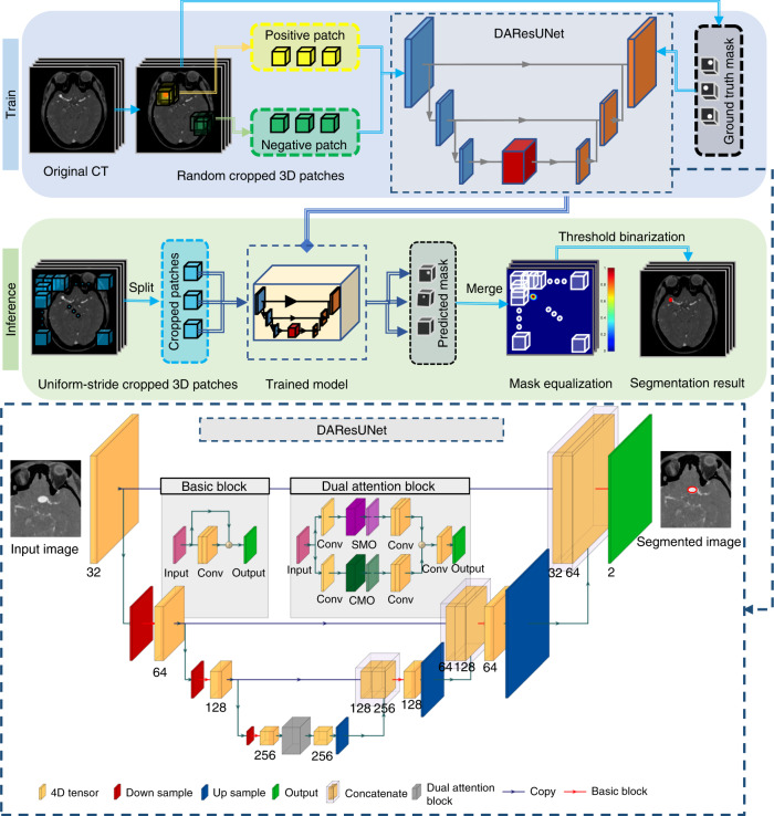
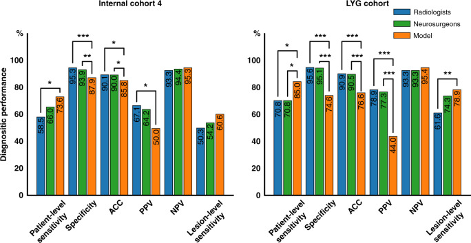
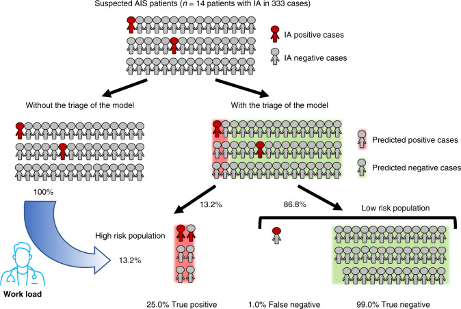
Intracranial aneurysm is a common life-threatening disease. Computed tomography angiography is recommended as the standard diagnosis tool; yet, interpretation can be time-consuming and challenging. We present a specific deep-learning-based model trained on 1,177 digital subtraction angiography verified bone-removal computed tomography angiography cases. The model has good tolerance to image quality and is tested with different manufacturers. Simulated real-world studies are conducted in consecutive internal and external cohorts, in which it achieves an improved patient-level sensitivity and lesion-level sensitivity compared to that of radiologists and expert neurosurgeons. A specific cohort of suspected acute ischemic stroke is employed and it is found that 99.0% predicted-negative cases can be trusted with high confidence, leading to a potential reduction in human workload. A prospective study is warranted to determine whether the algorithm could improve patients' care in comparison to clinicians' assessment.


Conflict of interest statement

Dr. Schoepf receives institutional research support from Bayer, Bracco, Guerbet, HeartFlow, Inc., and Siemens Healthineers and received personal fees for consulting and/or speaking from Bayer, Elucid BioImaging, General Electric, HeartFlow,. Inc., Keya Medical, and Siemens Healthineers. The other authors declare no competing interests.

Figures

Fig. 1
Fig. 1. Overview of the proposed DL model presented in this study.
a Training stage: we used 3D patches randomly sampled from the digital subtraction bone-removal CTA scans to train the network. b Inference stage: uniform-stride sampling was used and then the prediction of those samples was merged to obtain the final prediction of the whole volume. c Illustration of the architecture of the end-to-end aneurysm prediction model. The proposed segmentation model had a similar encoder–decoder architecture as U-Net, and residual blocks and a dual attention block were used to improve the performance of the network. IA intracranial aneurysm, SMO spatial matrix operation, CMO channel matrix operation.
Fig. 2
Fig. 2. Comparison of the performance of the model and the radiologists/neurosurgeons in Internal cohort 4 and LYG cohort.
A two-sided Pearson’s chi-squared test or Fisher exact test was used to evaluate the differences between the model and the radiologists and neurosurgeons. a Performance in Internal cohort 4. The model had higher patient-level sensitivity than that of the radiologists (χ2 = 4.337, p = 0.037) and comparative to neurosurgeons (χ2 = 0.934, p = 0.334). b Performance in external LYG cohort. Similar results can be found, i.e., that the model had a higher patient-level sensitivity than those of the radiologists (χ2 = 5.219, p = 0.022) and neurosurgeons (χ2 = 4.347, p = 0.037), and the specificity (χ2 = 140.346, p < 0.001 for the radiologists and χ2 = 69.381, p < 0.001 for the neurosurgeons), ACC (χ2 = 55.784, p < 0.001 for the radiologists and χ2 = 33.652, p < 0.001 for the neurosurgeons) and PPV (χ2 = 49.458, p < 0.001 for the radiologists and χ2 = 26.137, p < 0.001 for the neurosurgeons) were significantly lower for the model. ACC accuracy, NPV negative predictive value, PPV positive predictive value. *0.01 ≤ p < 0.05; **0.001 ≤ p < 0.01; ***p < 0.001.
Fig. 3
Fig. 3. Impact of the proposed model on clinical practice for patients with suspicion of AIS in the emergency department.
In the cohort of patients with suspected AIS (Internal cohort 5), who were prescribed to perform head CTA examination, 86.8% patients diagnosed as aneurysm-negative cases by our model, among which 99.0% were true-negative, demonstrating high confidence in identifying negative cases by our model. As a result, only 13.2% of patients were categorized as high-risk, to whom the radiologists can pay more-intense attention and reduce their workload in detecting aneurysm in AIS patients. AIS acute ischemic stroke, IA intracranial aneurysm.

References

    1. Vlak MH, et al. Prevalence of unruptured intracranial aneurysms, with emphasis on sex, age, comorbidity, country, and time period: a systematic review and meta-analysis. Lancet Neurol. 2011;10:626–636. doi: 10.1016/S1474-4422(11)70109-0. - DOI - PubMed
    1. van, Gijn J, Kerr RS, Rinkelm GJ. Subarachnoid haemorrhage. Lancet. 2007;369:306–318. doi: 10.1016/S0140-6736(07)60153-6. - DOI - PubMed
    1. Rincon F, Rossenwasser RH, Dumont A. The epidemiology of admissions of nontraumatic subarachnoid hemorrhage in the United States. Neurosurgery. 2013;73:217–222. doi: 10.1227/01.neu.0000430290.93304.33. - DOI - PubMed
    1. Connolly ES, Jr, et al. Guidelines for the management of aneurysmal subarachnoid hemorrhage. Stroke. 2012;41:1711–1737. doi: 10.1161/STR.0b013e3182587839. - DOI - PubMed
    1. Taufique Z, et al. Predictors of poor quality of life 1 year after subarachnoid hemorrhage. Neurosurgery. 2016;78:256–264. doi: 10.1227/NEU.0000000000001042. - DOI - PubMed

Publication types