Review
. 2020 Dec;52(12):1879-1890.
doi: 10.1038/s12276-020-00532-4. Epub 2020 Dec 1.

Stress and the dopaminergic reward system

Affiliations
Review

Stress and the dopaminergic reward system

Ja-Hyun Baik. Exp Mol Med. 2020 Dec.

Abstract

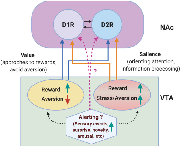
Dopamine regulates reward-related behavior through the mesolimbic dopaminergic pathway. Stress affects dopamine levels and dopaminergic neuronal activity in the mesolimbic dopamine system. Changes in mesolimbic dopaminergic neurotransmission are important for coping with stress, as they allow adaption to behavioral responses to various environmental stimuli. Upon stress exposure, modulation of the dopaminergic reward system is necessary for monitoring and selecting the optimal process for coping with stressful situations. Aversive stressful events may negatively regulate the dopaminergic reward system, perturbing reward sensitivity, which is closely associated with chronic stress-induced depression. The mesolimbic dopamine system is excited not only by reward but also by aversive stressful stimuli, which adds further intriguing complexity to the relationship between stress and the reward system. This review focuses on lines of evidence related to how stress, especially chronic stress, affects the mesolimbic dopamine system, and discusses the role of the dopaminergic reward system in chronic stress-induced depression.


Conflict of interest statement

The author declares no conflict of interest.

Figures

Fig. 1
Fig. 1. Dopaminergic pathways in the brain.
Schematic illustration of dopaminergic pathways in the central nervous system in the rodent brain. A8 cells are predominantly found in the retrorubral field (RRF), and A9 neurons are located in the substantia nigra pars compacta (SNc) and project to the dorsal striatum (DS), constituting the nigrostriatal pathway. The mesolimbic and mesocortical pathways include projections from A10 cells in the VTA to the nucleus accumbens (NAc), prefrontal cortex (PFC), and other limbic areas (which are not shown here). Other distinct groups of cells constituting the tuberoinfundibular pathway, namely, A12 cells and A14 cells in the hypothalamus, are shown.
Fig. 2
Fig. 2. Ventral tegmental area (VTA) and nucleus accumbens (NAc).
a Subareas within the VTA, in the lateral part of the VTA, the parabrachial pigmented area (PBP), which is continuous with the rostral SN (substantia nigra), and the paranigral nucleus (PN), which is rather restricted to the caudal VTA, are shown, and these regions are rich in DA neurons. The interfacial nucleus (IF) is found in the medial part of the VTA, the rostral linear nucleus of the raphe (RLi) is located in the rostral part of the VTA, and the caudal linear nucleus (CLi) is found in the caudal part of the VTA. The medial (Me) and lateral (L) parts of the VTA are indicated. b Schematic illustration of the NAc. The core and the shell (the medial and lateral parts of the shell are represented by (Me) and (L), respectively) of the NAc are showed together with the ventral pallidum (VP). DAergic neurons in the medial VTA (IF, PN, and medial PBP), which are schematically represented here by green projections, selectively project to the medial shell and core of the NAc, while DAergic neurons in the lateral VTA (lateral PBP), represented as pink projections, project to the lateral (L) shell of the NAc.
Fig. 3
Fig. 3. Reward and stress/aversion signals in the VTA-NAc pathway.
Based on recent findings, a diverse population of DAergic neurons that are excited (upward green arrow) by reward and inhibited (downward red arrow) by aversive stimuli and another population excited by both reward and aversive events are present in the VTA,,. In addition to being regulated by reward and aversive stimuli, DAergic neurons can be excited by numerous alerting signals (sensory events, surprise, novelty, arousal, attention, and salience), which do not necessarily associated have reward value. These value, salience, and alerting signals can cooperate to coordinate and control motivated behavior and may ultimately be important not only for reward value but also for supporting specific forms of adaptive behavior to react and cope with changes in the environment. These different DAergic neuronal populations may activate DAergic receptors in the NAc, but this process remains to be elucidated; here, the core and the shell of the NAc were not depicted. Two types of GABAergic medium spiny neuron (MSN) populations (90–95%) that constitute the majority of NAc neurons and express either dopamine D1R (D1-MSNs) or D2R (D2-MSNs) are represented as D1R and D2R, and their mutual connections are depicted by arrows. Other limbic areas are not described in the present figure.

References

    1. Selye H. A Syndrome produced by diverse nocuous agents. Nature. 1936;138:32. - PubMed
    1. Selye H. The evolution of the stress concept. Am. Sci. 1973;61:692–699. - PubMed
    1. McEwen BS. Stress, adaptation, and disease. Allostasis and allostatic load. Ann. N. Y. Acad. Sci. 1998;840:33–44. - PubMed
    1. Hammen C. Stress and depression. Ann. Rev. Clin. Psychol. 2005;1:293–319. - PubMed
    1. Pani L, Porcella A, Gessa G. The role of stress in the pathophysiology of the dopaminergic system. Mol. Psychiatry. 2000;5:14–21. - PubMed

Publication types