

Characteristics of secondary epiretinal membrane due to peripheral break
- PMID: 33257768
- PMCID: PMC7705695
- DOI: 10.1038/s41598-020-78093-9
Characteristics of secondary epiretinal membrane due to peripheral break
Abstract
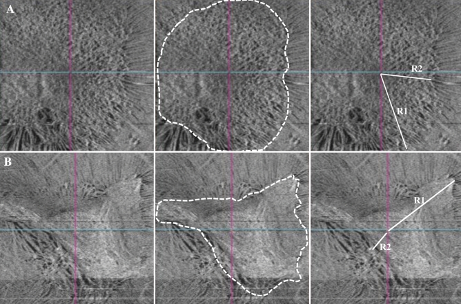
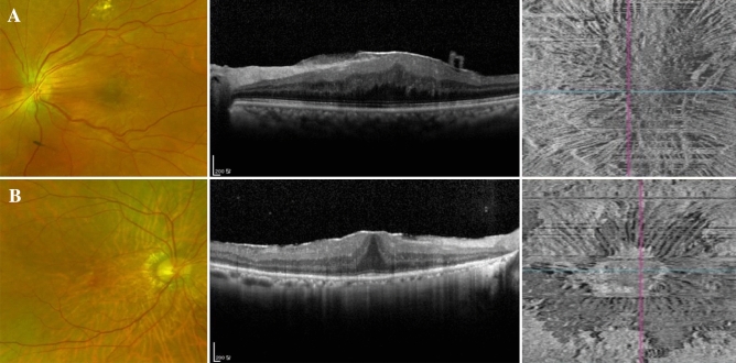
This study aimed to investigate morphological differences between idiopathic epiretinal membrane (ERM) and secondary ERM due to peripheral break (SEPB) and to identify clinical characteristics in eyes with SEPB to facilitate peripheral retinal examination. The retrospective cross-sectional study involved 93 consecutive eyes in 91 patients who underwent ERM removal surgery. Eyes were divided into two groups: the macular pucker group and the idiopathic ERM group. En-face Optical Coherence Tomography (OCT) images, fundus photographs, severity of metamorphopsia (M-score) and clinical characteristics of each group were compared. ERM extent and eccentricity (ratio of the shortest and longest distances from the foveal center to the boundary) were obtained through en-face OCT imaging. Fundus photographs were used to judge whether the membrane was turbid or not. Patients with SEPB were younger than patients with idiopathic ERM (61.3 ± 7.5 vs. 66.6 ± 8.3 years; p < 0.05). Preoperative M-score and myopic refractive error, axial length were also significantly higher in the macular pucker group than in the idiopathic ERM group (all p < 0.05). There was no difference in ERM extent between the two groups. The incidence of ERM eccentricity was 23 of the 34 eyes (67.6%) in the SEPB group and 26 of the 59 eyes (44.1%) in the idiopathic ERM group (p < 0.05). The incidence of turbid ERM was 18 of the 34 eyes (52.9%) in the SEPB group and 10 of the 59 eyes (16.9%) in the idiopathic ERM group (p < 0.01). The SEPB group, compared with the idiopathic ERM group, tended to have eccentric, turbid ERM at a younger age and with more severe metamorphopsia and myopic refractive error.
Conflict of interest statement
The authors declare no competing interests.
Figures
Similar articles
-
Influence of retinal vessel printings on metamorphopsia and retinal architectural abnormalities in eyes with idiopathic macular epiretinal membrane.Invest Ophthalmol Vis Sci. 2013 Nov 21;54(12):7803-11. doi: 10.1167/iovs.13-12817. Invest Ophthalmol Vis Sci. 2013. PMID: 24204051
-
THE EFFECT OF INTERNAL LIMITING MEMBRANE PEELING ON IDIOPATHIC EPIRETINAL MEMBRANE SURGERY, WITH A REVIEW OF THE LITERATURE.Retina. 2017 May;37(5):873-880. doi: 10.1097/IAE.0000000000001263. Retina. 2017. PMID: 27617536 Review.
-
Topographic changes in macula and its association with visual outcomes in idiopathic epiretinal membrane surgery.PLoS One. 2025 Jan 9;20(1):e0316847. doi: 10.1371/journal.pone.0316847. eCollection 2025. PLoS One. 2025. PMID: 39787120 Free PMC article.
-
Comparative analysis of metamorphopsia and aniseikonia after vitrectomy for epiretinal membrane, macular hole, or rhegmatogenous retinal detachment.PLoS One. 2020 May 8;15(5):e0232758. doi: 10.1371/journal.pone.0232758. eCollection 2020. PLoS One. 2020. PMID: 32384099 Free PMC article.
-
[Epiretinal membrane: diagnostics, indications and surgical treatment].Ophthalmologie. 2024 Jun;121(6):443-451. doi: 10.1007/s00347-024-02055-z. Epub 2024 Jun 3. Ophthalmologie. 2024. PMID: 38831204 Review. German.
Cited by
-
Potential of autophagy in subretinal fibrosis in neovascular age-related macular degeneration.Cell Mol Biol Lett. 2025 Apr 30;30(1):54. doi: 10.1186/s11658-025-00732-8. Cell Mol Biol Lett. 2025. PMID: 40307700 Free PMC article. Review.
-
Comparison of surgical outcomes after removal of epiretinal membrane associated with retinal break and idiopathic epiretinal membrane.Graefes Arch Clin Exp Ophthalmol. 2022 Jul;260(7):2121-2128. doi: 10.1007/s00417-021-05550-0. Epub 2022 Jan 14. Graefes Arch Clin Exp Ophthalmol. 2022. PMID: 35029729
-
Emerging clinical evidence of a dual role for Ang-2 and VEGF-A blockade with faricimab in retinal diseases.Graefes Arch Clin Exp Ophthalmol. 2025 May;263(5):1239-1247. doi: 10.1007/s00417-024-06695-4. Epub 2024 Dec 21. Graefes Arch Clin Exp Ophthalmol. 2025. PMID: 39708087 Free PMC article. Review.
-
Comparative electron microscopy analysis of internal limiting membrane and epiretinal membrane ultrastructure from vitrectomy surgery: A study protocol.PLoS One. 2025 Sep 2;20(9):e0331248. doi: 10.1371/journal.pone.0331248. eCollection 2025. PLoS One. 2025. PMID: 40892805 Free PMC article.
-
Optical Coherence Tomography Characteristics Between Idiopathic Epiretinal Membranes and Secondary Epiretinal Membranes due to Peripheral Retinal Hole.J Ophthalmol. 2025 May 7;2025:9299651. doi: 10.1155/joph/9299651. eCollection 2025. J Ophthalmol. 2025. PMID: 40371012 Free PMC article.
References
Publication types
MeSH terms
LinkOut - more resources
Full Text Sources
Research Materials
