. 2021 Feb;6(2):187-195.
doi: 10.1038/s41564-020-00815-6. Epub 2020 Nov 30.

A DNA-based vaccine protects against Crimean-Congo haemorrhagic fever virus disease in a Cynomolgus macaque model

Affiliations

A DNA-based vaccine protects against Crimean-Congo haemorrhagic fever virus disease in a Cynomolgus macaque model

David W Hawman et al. Nat Microbiol. 2021 Feb.

Abstract

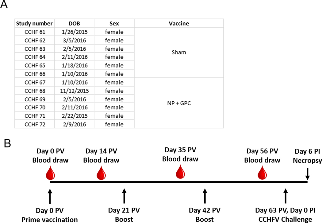
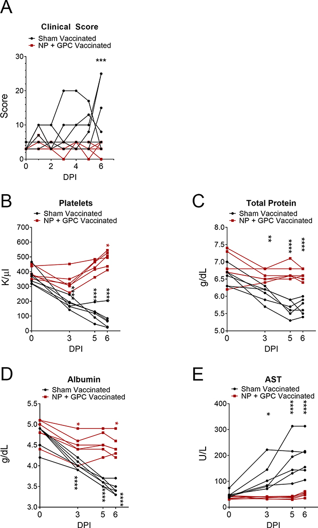
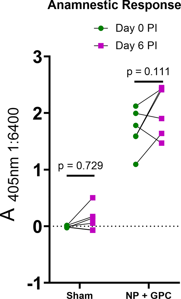
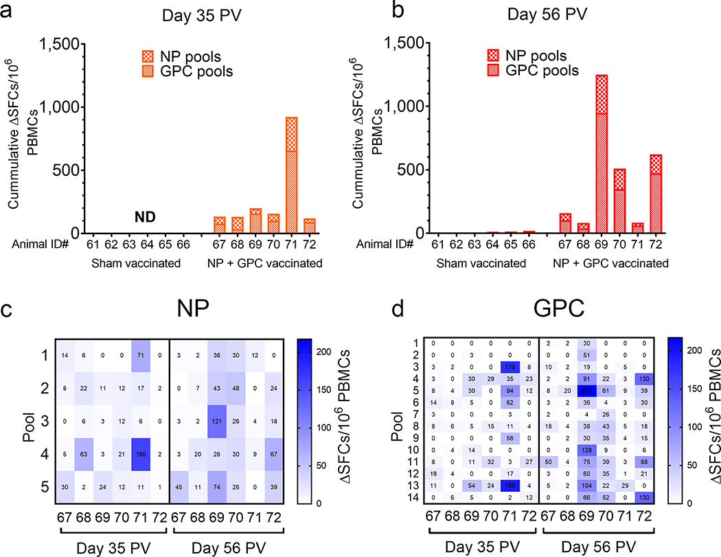
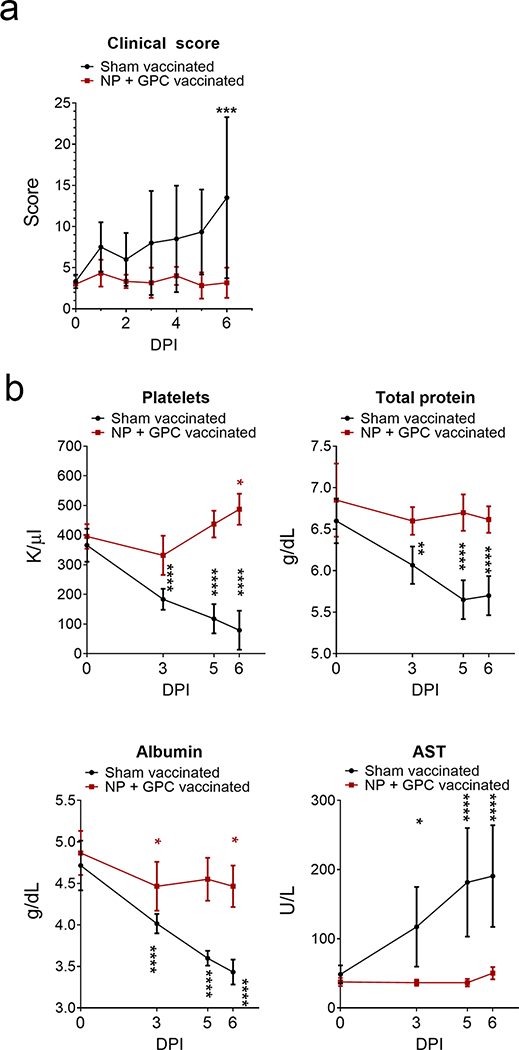
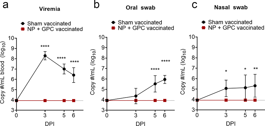
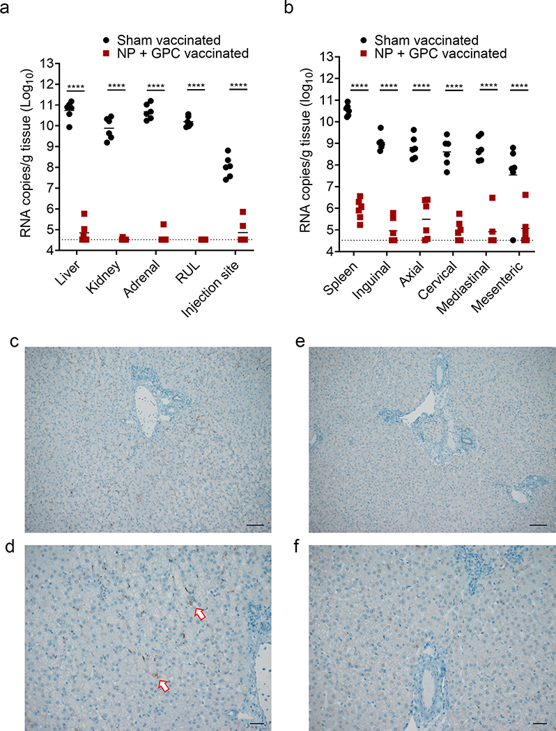
There is currently no specific prophylaxis or vaccine against Crimean-Congo haemorrhagic fever virus (CCHFV). Crimean-Congo haemorrhagic fever (CCHF) is a severe febrile illness transmitted by Hyalomma ticks in endemic areas, handling of infected livestock or care of infected patients. We report here the successful protection against CCHFV-mediated disease in a non-human primate disease model. Cynomolgus macaques were vaccinated with a DNA-based vaccine using in vivo electroporation-assisted delivery. The vaccine contained two plasmids encoding the glycoprotein precursor (GPC) and the nucleoprotein (NP) of CCHFV. Animals received three vaccinations and we recorded potent antibody and T cell responses after vaccination. While all sham-vaccinated animals developed viraemia, high tissue viral loads and CCHF-induced disease, the NP + GPC vaccinated animals were significantly protected. In conclusion, this is evidence of a vaccine that can protect against CCHFV-induced disease in a non-human primate model. This supports clinical development of the vaccine to protect groups at risk for contracting the infection.


Conflict of interest statement

Competing Interests

MS is a founder and chairman of the board of Svenska Vaccinfabriken Produktion AB.

Figures

Extended Data Fig. 1
Extended Data Fig. 1. Animal information and vaccination schedule
Animal information and vaccination schedule. (A) Animal number, date of birth (DOB), sex and vaccination. Animals were randomly assigned to either group prior to start of study. (B) Vaccination, blood draw and CCHFV challenge schedule. PV = post prime-vaccination, PI = post-CCHFV infection
Extended Data Fig. 2
Extended Data Fig. 2. Vaccination Site Histology
Vaccination Site Histology. At Day +6 CCHFV challenge, 27 days after last vaccination, tissue from the vaccination site was fixed in 10% formalin and stained with hematoxylin and eosin. Slides were scored by a pathologist (A). 0 = absent, 1 = mild, 2 = moderate, 3 = severe. (B-D) Representative images of the one severe (B), one moderate (C) and one of six mild animals (D) are shown at 100x magnification.
Extended Data Fig. 3
Extended Data Fig. 3. CCHFV ELISA on serum from sham-vaccinated animals
CCHFV ELISA on serum from sham-vaccinated animals. At indicated time points, serum was collected from sham-vaccinated animals and CCHFV-specific IgG measured by ELISA on day 0 or 56 post-prime vaccination. N = 6 animals per timepoint. Data shown as mean plus standard deviation.
Extended Data Fig. 4
Extended Data Fig. 4. Sandwich ELISA
Sandwich ELISA. A sandwich ELISA was performed on NP + GPC vaccinated animals to measure NP or Gc specific responses at day 0 post-prime vaccination (PV) and day 56 PV. N = 6 animals per timepoint. Statistical comparison performed using a two-way ANOVA with Sidak’s multiple comparison test.
Extended Data Fig. 5
Extended Data Fig. 5. Individual data for clinical scores and blood chemistry
Individual data for clinical scores and blood chemistry. The data from figure 3 is shown again but with individual data points shown.
Extended Data Fig. 6
Extended Data Fig. 6. Anamnestic response to CCHFV challenge
Anamnestic response to CCHFV challenge. A whole-virion ELISA was used to measure antibody responses in animals after CCHFV challenge on day 6 post-infection (PI). CCHFV-specific IgG was quantified in a 1:6400 dilution of serum. Statistical tests performed using a two-way ANOVA with Sidak’s multiple comparison test.
Extended Data Fig. 7
General plasmid schematics of plasmids used in this study.
Figure 1:
Figure 1:. NP + GPC vaccination induces robust antibody responses to CCHFV.
(A & B) At indicated time-points, CCHFV-specific IgM and IgG antibody responses in the serum were measured by whole-virion ELISA. Data is shown as the mean plus the standard deviation of duplicate measurements. Connecting lines were derived from unconstrained non-linear regression. Dashed line indicates average absorbance of background wells receiving no serum. (C) FRNT was performed to measure neutralizing activity of day −7 serum from NP + GPC vaccinated animals. Individual neutralization curves for each NP + GPC vaccinated animals are shown. Non-linear regression with a top constraint of 100 and bottom constraint of 0 was calculated and the FRNT50 determined. Data shown as mean plus standard error of measurement from triplicate technical replicates. N = 6 animals per group.
Figure 2:
Figure 2:. NP + GPC vaccination induces CCHFV-specific IFNγ T-cell responses.
Cryopreserved PBMCs from day −28 (A) or day −7 (B) were stimulated with pooled overlapping peptides derived from the CCHFV GPC or NP at 1μg/mL per peptide. Background was ascertained by wells stimulated with DMSO-vehicle alone. After 20 hours, IFNγ producing cells were detected by ELISpot. Spot forming cells (SFCs) were counted, background count subtracted from peptide-stimulated wells (ΔSFCs) and normalized to 1×10⁶ PBMCs. All measurements were performed in duplicate for each animal. (A & B) Data is shown as the sum of SFCs counted among all NP and GPC peptide pools. (C & D) For NP + GPC vaccinated animals the number of SFCs against each peptide pool for NP (C) and GPC (D) is shown. ND = not done.
Figure 3:
Figure 3:. NP + GPC vaccination improves clinical scores and blood chemistry following CCHFV challenge.
(A) Animals were comprehensively scored for evaluation of overt clinical disease and cumulative clinical scores are shown. Data shown as mean plus standard deviation. Statistical comparison was performed by two-way ANOVA with Sidak’s multiple comparison test at each time point. Day 6 PI P value = 0.0003. (B) At indicated time points platelets were enumerated in EDTA-treated whole-blood (normal range 300 – 500k/uL) and serum was evaluated for total protein (normal range 7.5 g/dL ±0.57), albumin (4.2 g/dL ±0.28) and aspartate aminotransferase (AST) (48.2IU/L ±36). Data is shown as mean plus standard deviation. Statistical comparison was performed between baseline values on day 0 and indicated time-point. N = 6 animals per group. Platelets, **** P < 0.0001, * P = 0.0129. Total Protein, **** P < 0.0001, ** P = 0.0027. Albumin, **** P < 0.0001, * P = 0.0131. AST, **** P < 0.0001, * P = 0.0280. P-values calculated with a two-tailed two-way ANOVA with Sidak’s multiple comparison test.
Figure 4:
Figure 4:. NP + GPC vaccination prevents viremia and viral shedding.
(A – C) At indicated time-points viral genomes were quantified by qRT-PCR. Data is shown as mean plus standard deviation. Dashed line indicates limit of detection. Viremia and Oral swab, **** p < 0.0001. Nasal swab, * P = 0.0169 (3 DPI) or 0.0109 (5 DPI), ** P = 0.0025. P-values calculated with a two-tailed two-way ANOVA with Sidak’s multiple comparison test. N = 6 animals per group.
Figure 5:
Figure 5:. NP + GPC vaccination reduces viral burden in multiple tissues.
(A & B) At day +6 PI a scheduled necropsy was performed, and viral genomes quantified in the indicated tissues by qRT-PCR. RUL = right upper lung. Individual values are shown, bar indicates mean and dashed line indicates limit of detection. **** p < 0.0001. P-values calculated with a two-tailed two-way ANOVA with Sidak’s multiple comparison test. (C – F) Immunohistochemistry to detect CCHFV antigen in the liver was performed and representative images of sham vaccinated (C & D) and NP + GPC vaccinated (E & F) animals are shown at 100x (C & E) or 200x (D & F) magnification. Arrows indicate antigen positive cells. Six sections from six animals per group were stained for CCHFV antigen and representative images from one animal per group shown. Scale bars indicate 50μm (C & E) or 100μm (D & F).

Similar articles

Cited by

References

    1. Bente DA et al. Crimean-Congo hemorrhagic fever: history, epidemiology, pathogenesis, clinical syndrome and genetic diversity. Antiviral Res 100, 159–189, doi:10.1016/j.antiviral.2013.07.006 (2013). - DOI - PubMed
    1. Negredo A et al. Autochthonous Crimean–Congo Hemorrhagic Fever in Spain. New England Journal of Medicine 377, 154–161, doi:10.1056/NEJMoa1615162 (2017). - DOI - PubMed
    1. Ergonul O Crimean-Congo haemorrhagic fever. Lancet Infect Dis 6, doi:10.1016/s1473-3099(06)70435-2 (2006). - DOI - PMC - PubMed
    1. Stuart MC, Kouimtzi M & Hill SR WHO Model Formulary 2008. (World Health Organization, 2008).
    1. Johnson S et al. Ribavirin for treating Crimean Congo haemorrhagic fever. The Cochrane database of systematic reviews 6, CD012713–CD012713, doi:10.1002/14651858.CD012713.pub2 (2018). - DOI - PMC - PubMed

Publication types

MeSH terms