. 2021 Feb;13(2):172-181.
doi: 10.1038/s41557-020-00586-x. Epub 2020 Nov 30.

Protein folding modulates the chemical reactivity of a Gram-positive adhesin

Affiliations

Protein folding modulates the chemical reactivity of a Gram-positive adhesin

Alvaro Alonso-Caballero et al. Nat Chem. 2021 Feb.

Abstract

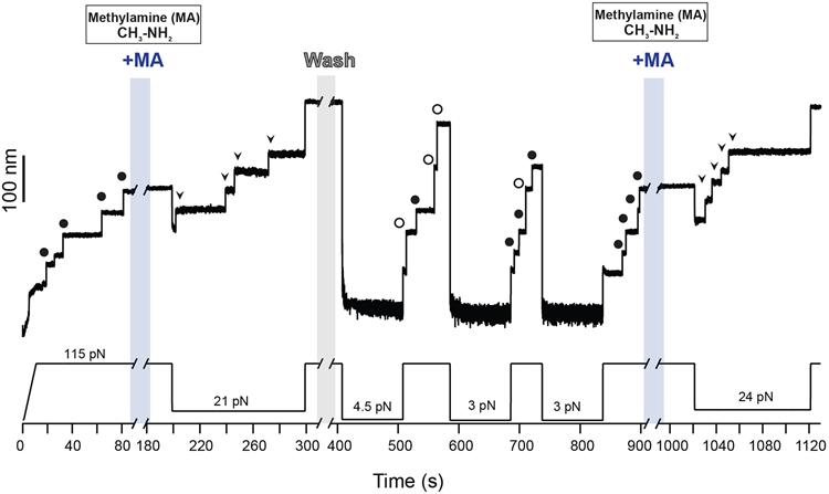
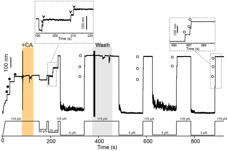
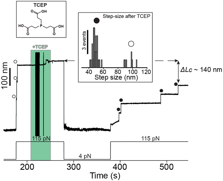
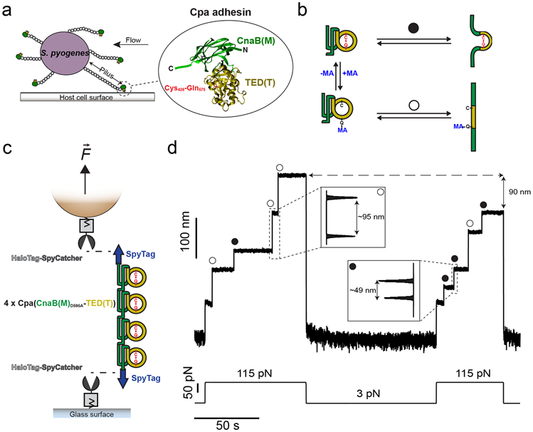
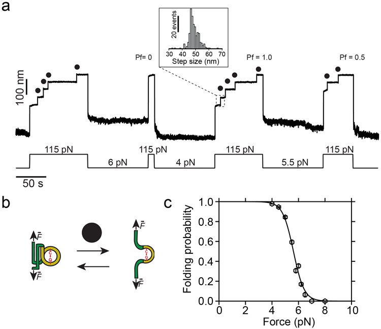
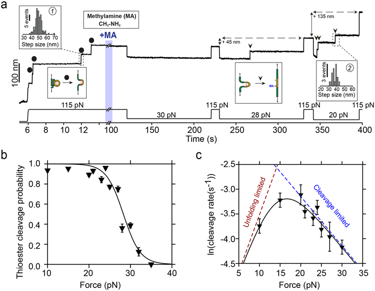
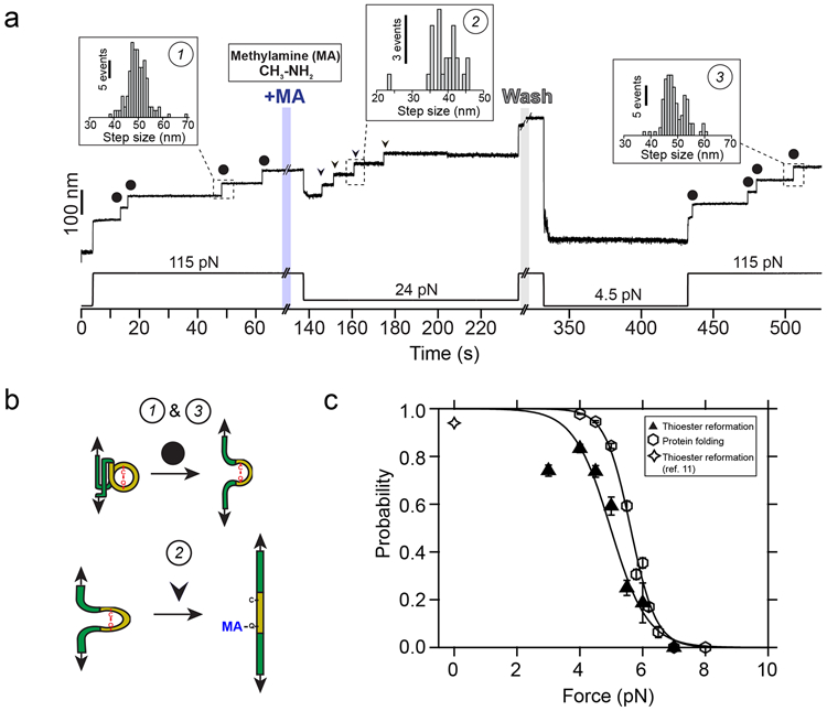
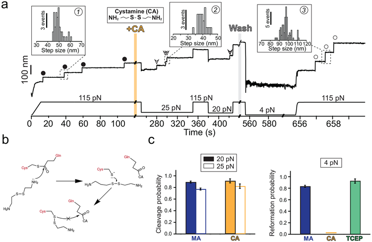
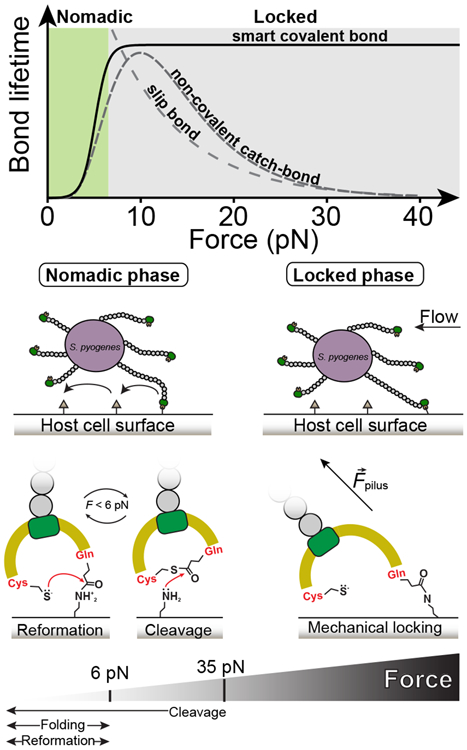
Gram-positive bacteria colonize mucosal tissues, withstanding large mechanical perturbations such as coughing, which generate shear forces that exceed the ability of non-covalent bonds to remain attached. To overcome these challenges, the pathogen Streptococcus pyogenes utilizes the protein Cpa, a pilus tip-end adhesin equipped with a Cys-Gln thioester bond. The reactivity of this bond towards host surface ligands enables covalent anchoring; however, colonization also requires cell migration and spreading over surfaces. The molecular mechanisms underlying these seemingly incompatible requirements remain unknown. Here we demonstrate a magnetic tweezers force spectroscopy assay that resolves the dynamics of the Cpa thioester bond under force. When folded at forces <6 pN, the Cpa thioester bond reacts reversibly with amine ligands, which are common in inflammation sites; however, mechanical unfolding and exposure to forces >6 pN block thioester reformation. We hypothesize that this folding-coupled reactivity switch (termed a smart covalent bond) could allow the adhesin to undergo binding and unbinding to surface ligands under low force and remain covalently attached under mechanical stress.


Figures

Extended Data Fig. 1
Extended Data Fig. 1. Cleavage-reformation-cleavage sequence
Magnetic tweezers force-clamp trajectory of the Cpa polyprotein. After the unfolding of four thioester-intact Cpa domains at 115 pN (circles, ~49 nm), the buffer is exchanged and the polyprotein is exposed to a solution containing 100 mM methylamine (+MA). At 21 pN, four steps appear which account for the release of the polypeptide sequence trapped by the thioester bonds (arrows). Then, the force is increased again to 115 pN, revealing the complete extension of the molecule. Immediately after, MA is washed out from the fluid chamber and the polyprotein is allowed to fold and reform the thioester bonds for 100 s at 4.5 pN. A 115 pN pulse reveals three ~95 nm steps (empty circles) which correspond with the full extension of Cpa, and one Cpa domain with its thioester bond reformed (circles, ~49 nm). Two more quenches at 3 pN are applied to completely recover the thioester-reformed state in all the four domains, as it can be seen in the 115 pN pulse applied approximately after 800 s of experiment (circles). Then, MA is added again and the force quenched to 24 pN to trigger again the cleavage of the thioester bonds of the polyprotein (arrows).
Extended Data Fig. 2
Extended Data Fig. 2. Cystamine permanent blocking of Cpa thioester bond reformation
Magnetic tweezers force-clamp trajectory of the Cpa polyprotein. After the unfolding of the thioester-intact Cpa domains at 115 pN (circles), the buffer is exchanged and the polyprotein is exposed to a solution containing 100 mM cystamine (+CA). At 115 pN and at 50 pN, no additional extensions are registered as a consequence of thioester bond cleavage, but a drop in force to 25 pN leads to the appearance of four steps which account for the release of the polypeptide sequence trapped by the thioester bonds (empty arrows in the inset). Then, the force is increased again to 115 pN, revealing the complete extension of the molecule. After 100 s at 4 pN and in the presence of CA, a 115 pN pulse reveals three ~95 nm steps (empty circles) which correspond with the full extension of Cpa. CA is then removed from the solution, and several consecutive 100 s force quenches (at 4, 5, and 3 pN) followed by 115 pN pulses are applied. These cycles reveal that, after CA treatment, Cpa is able to fold but not to reform its thioester bond, as it can be observed from the ~95 nm steps observed (empty circles). After the first 300 s of the experiment, one of the Cpa domains stops folding back as a consequence of oxidative damage. The disturbances observed in the extension during +CA addition (orange block) and washing (gray block) are originated from the movement of buffer volumes in the liquid cell used in the experiments, which transiently alter the measurement
Extended Data Fig. 3
Extended Data Fig. 3. TCEP rescues Cpa thioester bond reformation
A Cpa polyprotein previously treated with cystamine shows three ~95 nm steps at 115 pN corresponding with the full extension of each of the domains (empty circles). The addition of 10 mM TCEP and 100 s at 4 pN is enough to trigger thioester bond reformation, as it can be observed in the ~49 nm thioester-intact Cpa steps (circles) registered at 115 pN. The fourth domain not observed at the beginning was probably unfolded and its thioester bond intact, since the difference in the final extension between the first 115 pN pulse and the last is ~140 nm, which matches with the expected final extension decrease from three reformation events. Inset histogram shows the two populations of steps observed after TCEP treatment, thioester-intact Cpa (circles, 48.3 ± 3.5 nm, mean±SD, n=32) and thioester-cleaved Cpa (empty circles, 95.7 ± 6.4 nm mean±SD, n=7). The latter full length steps of Cpa after TCEP treatment could be due to cleavage events induced by remaining cystamine which was not completely washed from the experimental liquid cell. The disturbances observed in the extension during +TCEP addition (green block) are originated from the movement of buffer volumes in the liquid cell used in the experiments, which transiently alter the measurement.
Figure 1.
Figure 1.. Mechano-chemistry of S. pyogenes Cpa adhesin.
a) S. pyogenes attach to host cell surfaces through the Cpa protein, present in the tip-end of the pili. Cpa main core comprises the CnaB(M) domain (green), in whose fold the TED(T) domain is inserted (yellow). The TED(T) domain contains a thioester bond formed between the residues Cys426 and Gln575 (red), which mediates the attachment to cell-surface molecules. b) In the folded state, nucleophiles like methylamine (MA) can cleave the thioester bond and bind covalently to the Gln side chain (+MA); however, thioester bond reformation and ligand uncoupling (−MA) can occur. After mechanical extension, the presence (circle pathway) or absence (empty circle pathway) of the thioester bond can be assessed as a difference in the extension of the protein. c) Double-covalent magnetic tweezers experimental assay. Protein anchors SpyCatcher-HaloTag are covalently immobilized both to the surface of the glass and the paramagnetic bead. A chimeric polyprotein made of four copies of Cpa and flanked by SpyTag peptides is covalently linked to the glass and the bead through the reaction of the SpyCatcher/SpyTag split protein system. On the top of the scheme (not shown), the position of a pair of magnets is controlled for the application of calibrated forces to the tethered molecule. d) Magnetic tweezers recording of a Cpa polyprotein exposed to 100 mM methylamine, where the extension of the molecule is registered along time. A force pulse of 115 pN leads to the mechanical unfolding of the four Cpa domains, which is detected as stepwise increases in the extension. Here, three of the domains lack their internal thioester bond (empty circles) yielding an extension of ~95 nm, while one of the domains preserves its thioester bond (circle) and yields an unfolding extension of ~49 nm. Following a 100 s-long quench force pulse at 3 pN, which favors both folding and bond reformation, a second 115 pN pulse reveals that two Cpa domains reformed their thioester bonds (circles), decreasing the final extension of the polyprotein by 90 nm, as a consequence of the polypeptide sequence trapped by the newly formed bonds.
Figure 2.
Figure 2.. Dynamics of the thioester-intact Cpa polyprotein under force.
a) Magnetic tweezers trajectory of the Cpa polyprotein. High force pulses at 115 pN unfold the thioester-intact Cpa domains, which show 48.8 ± 3.8 nm (mean±SD, n=272) stepwise extensions (inset histogram). Low force pulses of 100 seconds long allow Cpa refolding, enabling us to determine the folding probability (Pf) at different forces. As an example, a quench at 6 pN does not allow folding of any of the domains, while the four-fold at 4 pN (Pf=1.0), and only two-fold at 5.5 pN (Pf=0.5). b) Cartoon representation of the folding-unfolding of the Cpa domain. The thioester bond between Cys426 and Gln575 clamps the TED domain (yellow), limiting its extensibility. c) Folding probability of thioester-intact Cpa. Data points are fitted to a sigmoidal function and they represent the probability at each of the forces tested for 100 s (n=54 at 4 pN; n=30 at 4.5 pN; n=18 at 5 pN; n=16 at 5.5 pN; n=16 at 5.8 pN; n=23 at 6 pN; n=14 at 6.2 pN; n=10 at 6.5 pN; n=9 at 7 pN; n=5 at 8 pN). Data points are the mean and the bars are the SD calculated using a jackknife analysis.
Figure 3.
Figure 3.. Cpa thioester bond cleavage is negatively force-dependent.
a) Magnetic tweezers trajectory of the Cpa polyprotein. After the unfolding of the thioester-intact Cpa domains at 115 pN (circles; histogram inset #1: 49.6±4.1 nm, mean±SD, n=164), the buffer is exchanged to a Hepes solution containing 100 mM methylamine (+MA). At high force, no additional steps are registered as it would be expected from a thioester bond cleavage event. Thereafter, we apply a protocol with subsequent pulses of decreasing mechanical load to investigate the force-dependency of the reaction. While at 30 pN no cleavage is observed, 100 s at 28 pN reveal one step that comes from the methylamine-induced cleavage of the thioester bond of one of the four Cpa domains (triangle). At 115 pN, the final extension of the molecule has increased by 45 nm, which originates from the polypeptide sequence released after thioester bond lysis. When held at 20 pN, the three remaining thioester bonds are cleaved (triangles; histogram inset #2: 38± 3.1 nm, mean±SD, n=21) and the final extension of the molecule increases for another 135 nm. b) Thioester bond cleavage probability as a function of force measured over a 100 s time-window. Data points are the mean and the error bars are the SD calculated using a jackknife analysis. The line represents a sigmoidal fit to the data (n=12 at 10 pN; n=20 at 15 pN; n=15 at 20 pN; n=9 at 21 pN; n=10 at 23 pN; n=9 at 24 pN; n=15 at 25 pN; n=15 at 27 pN; n=7 at 28 pN; n=15 at 30 pN; n=5 at 32 pN; n=6 at 35 pN). c) Rate of thioester bond cleavage as a function of force. Data points show the natural logarithm of the cleavage rate and the bars show the standard error of the mean. The curve represents a fit to the data described by a model that takes into account the effect of two sequential reactions: the rate of protein unfolding, which increases with force, and the rate of thioester bond cleavage, which decreases with the force. From this fit, we obtain a distance to the transition state for TED protein unfolding (xU) of 0.9 nm, while the thioester bond cleavage exhibits a negative distance to the transition state (xC = −0.4 nm), which suggests a requirement of a contraction of the Cpa polypeptide substrate to proceed with the cleavage of the bond, explaining its negative force-dependence. The dotted lines represent the individual unfolding and cleavage rates as obtained from the fit to the proposed model (Eq. S3, see Methods) (n=30 at 10 pN; n=38 at 15 pN; n=24 at 20 pN; n=23 at 21 pN; n=23 at 23 pN; n=24 at 24 pN; n=21 at 25 pN; n=37 at 27 pN; n=15 at 30 pN). Rate vs force dependency data was obtained in unrestricted time windows experiments.
Figure 4.
Figure 4.. Protein folding drives thioester bond reformation.
a) Magnetic tweezers trajectory of the Cpa polyprotein. After the unfolding of the thioester-intact Cpa domains at 115 pN (circles; inset histogram #1: 49.6 ± 4.1 nm, mean±SD, n=164), the buffer is exchanged and the polyprotein is exposed to a solution containing 100 mM methylamine (+MA). As expected, we do not observe cleavage at this high force, but a drop to 24 pN permits the full cleavage of the four candidate thioester bonds (arrows, inset histogram #2; 38.8 ± 4.4 nm for 24 pN, mean±SD, n=25). To study the reformation of the bond, we remove the nucleophile-containing buffer at high force, and quench the force to 4.5 pN for 100 s to favor bond reformation and protein folding. We stretch again the polyprotein at 115 pN and identify four thioester-intact Cpa domains, which indicates that the four cleaved candidates were able to fold and to reform their bonds (circles; inset histogram #3: 48.8 ± 4.1 nm, mean±SD, n=117). b) Cartoon representation of the extension events registered on the Cpa trajectory shown in a). Events #1 and #3 show the mechanical extension at 115 pN of thioester-intact Cpa, before cleavage and after reformation, respectively. Event #2 shows the extension after methylamine (MA) cleavage at 24 pN. c) Comparison between the thioester bond reformation (upwards triangles and sigmoidal fit) and the thioester-intact Cpa folding probability (hexagons and sigmoidal fit, from Figure 2c) as a function of the mechanical load. Star symbol indicates the reformation probability obtained at 0 pN from our previous work with AFM. Data points for reformation are the mean and the error bars are the SD calculated using a jackknife analysis. Reformation registered as the amount of thioester-intact domains after methylamine washout and after a 100 s time-window at the folding/reformation force range (n=13 at 3 pN; n=16 at 4 pN; n=15 at 4.5 pN; n=12 at 5 pN; n=6 at 5.5 pN; n=7 at 6 pN; n=6 at 7 pN).
Figure 5.
Figure 5.. Cystamine-mediated abrogation of Cpa thioester bond reformation.
a) Magnetic tweezers trajectory of the Cpa polyprotein. After the unfolding of the thioester-intact Cpa domains at 115 pN (circles, inset histogram #1; 49.3 ± 3.8 nm, mean±SD, n=42), the buffer is exchanged and the polyprotein is exposed to a solution containing 100 mM cystamine (+CA). At 115 pN, no additional extensions are registered, but a drop in the force to 25 pN for 100 s leads to the appearance of three steps which account for the release of the polypeptide sequence trapped by the thioester bonds (empty arrows, inset histogram #2; 39.6 ± 2.7 nm for 25 pN, mean±SD, n=23). After the cleavage of all the bonds and after CA washout, force is quenched to 4 pN for 100 s to favor folding and reformation of the thioester. The final 115 pN pulse reveals three steps corresponding to thioester bond-cleaved Cpa domains (empty circles, inset histogram #3; 97.1 ± 5.2 nm, mean±SD, n= 78). b) Chemical scheme depicting the reformation blocking effect of CA. After the thioester bond nucleophilic cleavage by one of the CA primary amines, the free Cys thiol can attack CA disulfide bond (from the bound CA, or from another CA molecule). As a result, an intermolecular disulfide bond between Cpa Cys426 and CA is formed, preventing the thioester bond reformation. This disulfide reshuffling breaks the CA molecule and generates one free CA molecule (not shown in the scheme), and a Cys426-bound CA. c) Left panel compares the thioester bond cleavage probability by methylamine (MA) and CA at 20 and at 25 pN (MA, n=15 at 20 pN, n=15 at 25 pN; CA, n=8 at 20 pN, n=9 at 25 pN). Right panel compares the thioester bond reformation probability after 100 s at 4 pN after the treatment with MA, CA, and after the treatment with CA followed by TCEP (MA, n=16; CA, n=17; TCEP, n=6). Histogram bars are the mean and the error bars are the SD calculated using a jackknife analysis
Figure 6.
Figure 6.. Bacterium mobility strategy model based on the allosteric modulation of the Cpa thioester bond by protein folding.
Top graph compares bond lifetimes as a function of the mechanical load for slip bonds, non-covalent catch bonds, and smart covalent bonds (slip bond and catch bond data adapted from, plotted in arbitrary units). The smart covalent bond lifetime (plotted as the inverse of the thioester bond reformation probability from Figure 4c) is defined as the lifetime of the bond made between the surface ligand and the Gln575 side chain after the nucleophilic cleavage of the thioester bond. While higher loads decrease exponentially the lifetime of slip bonds, in non-covalent catch bonds it increases; however, loads above certain threshold decrease the lifetime. The adhesin-ligand smart covalent bond is allosterically modulated by force, establishing short-lived bonds with surface ligands at low mechanical stress—where thioester bond reformation and cleavage coexist—when the protein is folded, but turning into a long-lived bond that permits the bacterium to remain attached under large mechanical challenges, where thioester bond reformation is prevented. We hypothesize that these smart covalent bonds could allow bacteria to switch between a nomadic mobility phase at low force to a mechanically locked phase at larger loads.

References

    1. Hall-Stoodley L, Costerton JW & Stoodley P Bacterial biofilms: from the Natural environment to infectious diseases. Nat. Rev. Microbiol 2, 95–108 (2004). - PubMed
    1. Yan J & Bassler BL Surviving as a Community: Antibiotic Tolerance and Persistence in Bacterial Biofilms. Cell Host Microbe 26, 15–21 (2019). - PMC - PubMed
    1. Forero M, Yakovenko O, Sokurenko EV, Thomas WE & Vogel V Uncoiling mechanics of Escherichia coli type I fimbriae are optimized for catch bonds. PLoS Biol. 4, 1509–1516 (2006). - PMC - PubMed
    1. Sauer MM et al. Catch-bond mechanism of the bacterial adhesin FimH. Nat. Commun 7, 10738 (2016). - PMC - PubMed
    1. Pointon JA et al. A Highly Unusual Thioester Bond in a Pilus Adhesin Is Required for Efficient Host Cell Interaction. J. Biol. Chem 285, 33858–33866 (2010). - PMC - PubMed

References (Methods)

    1. Valle-Orero J et al. Mechanical Deformation Accelerates Protein Ageing. Angew. Chemie Int. Ed 56, 9741–9746 (2017). - PMC - PubMed

Publication types

MeSH terms

Substances