

Case Report: Scabies Invading Gingival Tissue
- PMID: 33258442
- PMCID: PMC7790058
- DOI: 10.4269/ajtmh.20-0707
Case Report: Scabies Invading Gingival Tissue
Abstract
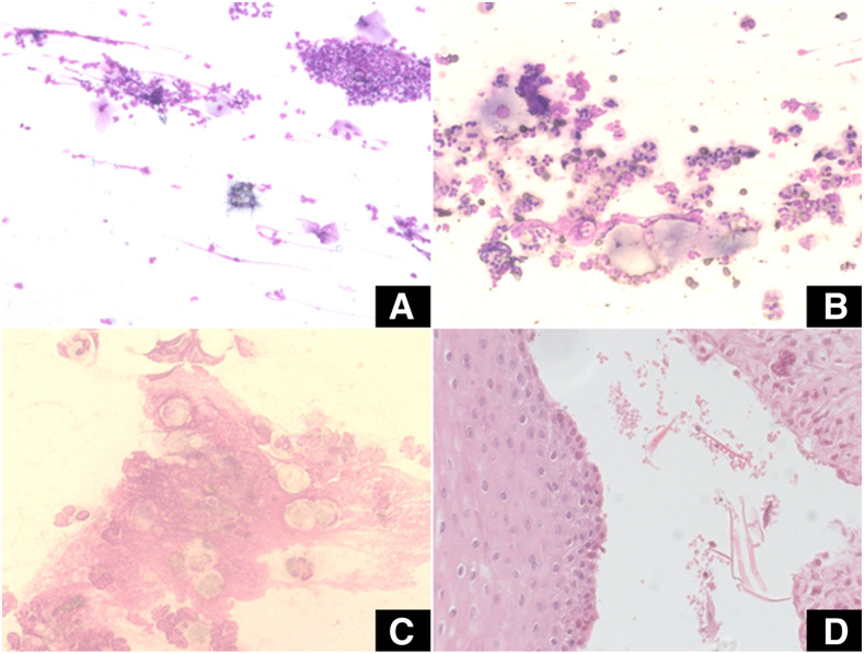
Non-plaque-induced lesions may occur on the gingiva as medical disorders or manifestations of systemic conditions. Scabies is a parasitic infection caused by Sarcoptes scabiei. Here, we present the first case of oral scabies reported in the literature located on the gingiva in a 43-year-old woman. She was admitted to the hospital complaining of an ulcerative lesion on the gingiva with unknown duration, with a suggestive diagnosis of pemphigoid. A diagnosis of scabies infestation was made based on the visualization of eggs and larvae/nymph forms. The treatment consisted of 100 mg of ivermectin (three times per day for 15 days), supplemental oral hygiene with chlorhexidine, and extensive cleaning. The follow-up was made 30 days after treatment with ivermectin. The patient did not report side effects, with skin and oral lesions completely healed. Based on this, we need to perform a thoughtful ectoscopy examination and be alert to signs that indicate unusual causes to diagnose correctly and choose the appropriate treatment.
Figures

References
Publication types
MeSH terms
Substances
LinkOut - more resources
Full Text Sources
Other Literature Sources
Medical
