. 2020 Dec 1;45(23):6378-6381.
doi: 10.1364/OL.404415.

Active line scan with spatial gating for sub-diffuse reflectance imaging of scatter microtexture

Active line scan with spatial gating for sub-diffuse reflectance imaging of scatter microtexture

Samuel S Streeter et al. Opt Lett. .

Abstract

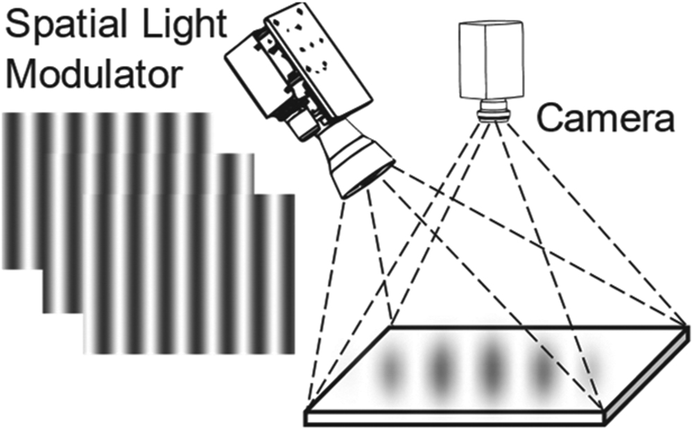
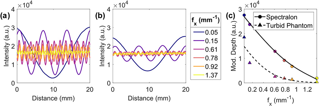
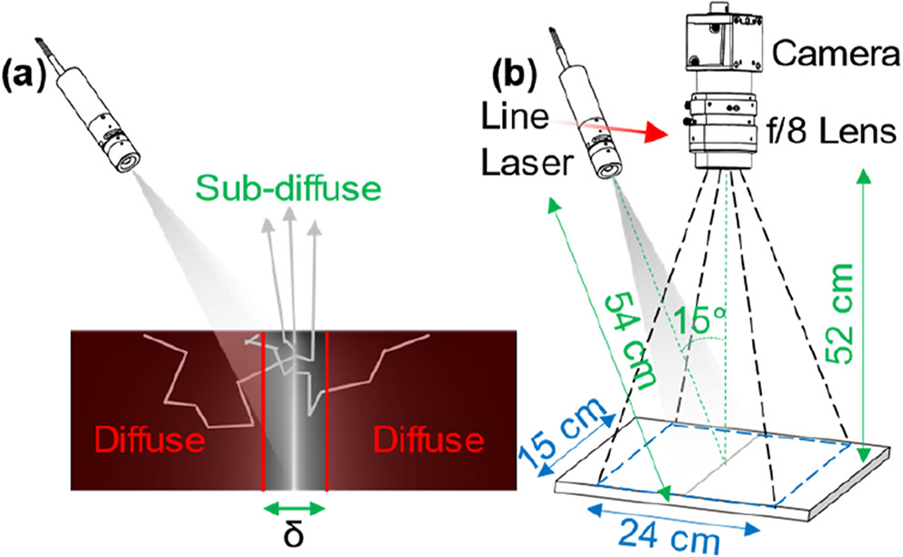
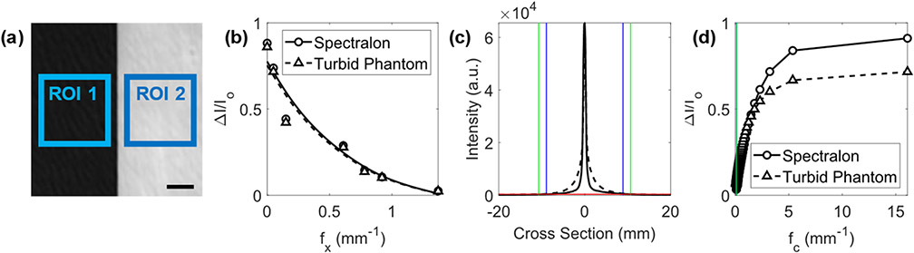
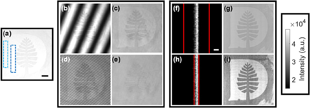
We examine the value of an active line scan with spatial gating for imaging sub-diffuse, wide-field reflectance microtexture. Line scanning combined with spatial gating and linear translation can be used for localized detection of features in the surface layer of a turbid target. The line scan provides broadband spatial frequency modulation, and the spatial gating effectively high-pass filters the reflectance. The major benefit of this approach is that of high dynamic range (70%-90%) signal preservation and high contrast to noise when imaging at high spatial frequencies. Alternative approaches, such as spatial frequency domain imaging, are degraded by low dynamic range in demodulated images, making it nearly impossible to image over a wide field of view at frequencies over 1.5mm-1 using commercial technology. As such, active line scanning with spatial gating presents as an inherently high sensitivity and high dynamic range method of imaging microscopic scattering features in only the surface layer of a turbid medium.


Conflict of interest statement

Disclosures. The authors declare no conflicts of interest.

Figures

Fig. 1.
Fig. 1.
Reflectance geometry SFDI, with images taken at three phases and demodulated to isolate reflectance from a specific spatial frequency and wavelength of light.
Fig. 2.
Fig. 2.
Measurements of SFDI modulation patterns at a single illumination phase before demodulation. Modulation patterns (λ = 600 nm) at six discrete spatial frequencies reflected from (a) Spectralon and (b) the turbid phantom. (c) Modulation depth of each pattern shown in (a) as circles and in (b) as triangles with fitted curves, becoming unusable ~fx > 1.5 mm−1.
Fig. 3.
Fig. 3.
(a) Laser line with a spatial gate width of δ isolates reflectance from predominantly sub-diffusely scattered photons. (b) Schematic of the line scanning setup used in this study.
Fig. 4.
Fig. 4.
(a) Black board (ROI 1) and Spectralon or turbid phantom (ROI 2) high contrast target with a 5 mm scale bar. Relative dynamic range quantified using (b) SFDI images with respect to fx (λ = 600 nm, shown with fitted curves) and (d) line scanning with respect to fc (λ = 410 nm). In (c), the noise floor of reflectance lines from the Spectralon and turbid phantom (red line) define the maximum gate width (Spectralon, blue lines, δ ≈ 18 mm; turbid phantom, green lines, δ ≈ 22 mm). Corresponding fc values are shown in (d) along left edge with the same coloring.
Fig. 5.
Fig. 5.
A Dartmouth College Pine logo target demonstrates sensitivity to scattering microtexture. (a) The target under room light with a 1 cm scale bar, showing the construction paper (dark blue) and painted (light blue) regions. (b) SFDI at fx = 0.05 mm−1 yields the demodulated reflectance image in (c). (d) SFDI at fx = 0.61 mm−1 yields the demodulated reflectance image in (e). (f) Close-up of a laser line with δ = 5 mm (red lines) and a 1 mm scale bar and (g) corresponding demodulated reflectance image. (h) Close-up of a laser line with δ = 1 mm and (i) corresponding demodulated reflectance image. All images are shown without histogram stretching.

References

    1. Neil MAA, Juškaitis R, and Wilson T, Opt. Lett 22, 1905 (1997). - PubMed
    1. Wilson T, J. Microsc 242, 111 (2011). - PubMed
    1. Gustafsson MGL, J. Microsc 198, 82 (2000). - PubMed
    1. Geng J, Adv. Opt. Photon 3, 128 (2011).
    1. Kanick SC, McClatchy DM, Krishnaswamy V, Elliott JT, Paulsen KD, and Pogue BW, Biomed. Opt. Express 5, 3376 (2014). - PMC - PubMed