. 2020 Dec;17(173):20200614.
doi: 10.1098/rsif.2020.0614. Epub 2020 Dec 2.

Biomimetic six-axis robots replicate human cardiac papillary muscle motion: pioneering the next generation of biomechanical heart simulator technology

Affiliations

Biomimetic six-axis robots replicate human cardiac papillary muscle motion: pioneering the next generation of biomechanical heart simulator technology

Annabel M Imbrie-Moore et al. J R Soc Interface. 2020 Dec.

Abstract

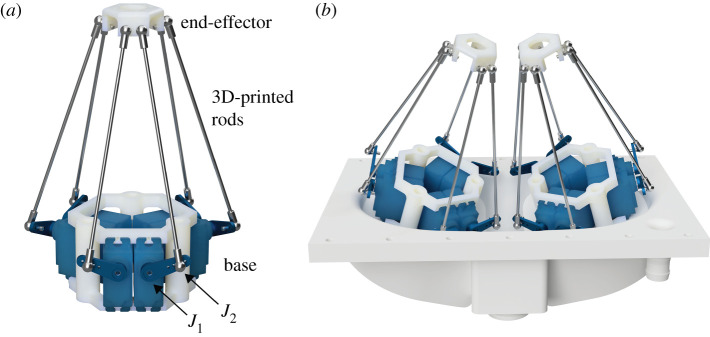
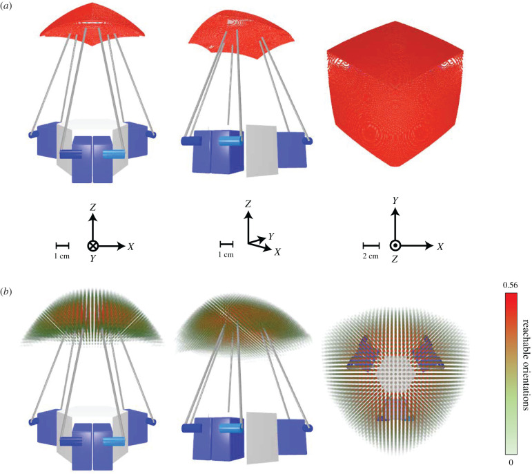
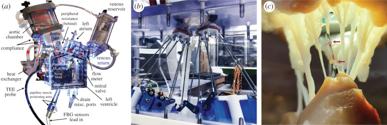
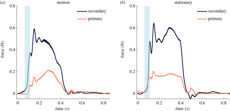
Papillary muscles serve as attachment points for chordae tendineae which anchor and position mitral valve leaflets for proper coaptation. As the ventricle contracts, the papillary muscles translate and rotate, impacting chordae and leaflet kinematics; this motion can be significantly affected in a diseased heart. In ex vivo heart simulation, an explanted valve is subjected to physiologic conditions and can be adapted to mimic a disease state, thus providing a valuable tool to quantitatively analyse biomechanics and optimize surgical valve repair. However, without the inclusion of papillary muscle motion, current simulators are limited in their ability to accurately replicate cardiac biomechanics. We developed and implemented image-guided papillary muscle (IPM) robots to mimic the precise motion of papillary muscles. The IPM robotic system was designed with six degrees of freedom to fully capture the native motion. Mathematical analysis was used to avoid singularity conditions, and a supercomputing cluster enabled the calculation of the system's reachable workspace. The IPM robots were implemented in our heart simulator with motion prescribed by high-resolution human computed tomography images, revealing that papillary muscle motion significantly impacts the chordae force profile. Our IPM robotic system represents a significant advancement for ex vivo simulation, enabling more reliable cardiac simulations and repair optimizations.

Keywords: biomechanics; cardiac imaging; ex vivo modelling; robotics.


Conflict of interest statement

We declare we have no competing interests.

Figures

Figure 1.
Figure 1.
Diagram of the IPM robot. (a) A single IPM robot. J1 denotes the first joint at the base of the servo arm and J2 denotes the second joint connecting the servo arm to the 3D-printed rod. (b) Render of the dual IPM robotic system mounted on the base plate of the left ventricular chamber in the heart simulator. The platforms are both angled 15° towards the centre for additional operating space.
Figure 2.
Figure 2.
Range-of-motion simulation results. Simulation results calculating the total range of motion of the IPM robot end-effector via inverse kinematics iteration on the Stanford Sherlock supercomputing cluster. Axes and scale for each view are indicated. (a) Three-dimensional iteration, with a fixed, neutral orientation, absent of rotation, and a resolution of 0.11 mm. (b) Six-dimensional iteration with a resolution of 3.33 mm and 3.75°. Greater opacity and red colour indicate a higher relative frequency of reachable orientations for a given position with a maximum reachable orientation frequency of 0.56. For reference, a relative frequency rate of reachable orientations of 1 indicates a position where every permutation of orientations with the bounds of [−π/2, π/2] for each axis is reachable. The dynamic range of the reachable orientation percentages was exponentially tuned for better visibility.
Figure 3.
Figure 3.
Heart simulator experimental setup. (a) Diagram of the custom left heart simulator with stationary papillary muscles. From Imbrie-Moore [8], used with permission from Elsevier. (b) Picture of the dual Stewart platform system sewn to papillary muscles of a porcine mitral valve to mimic the motion of the heart during ex vivo cardiac simulation experiments. (c) High-resolution FBG strain gauge sensor instrumenting a chordae tendineae; the sensor is calibrated to correlate strain to force. The chord is severed between the two suture attachment points, denoted by red arrows, to transmit all force through the sensor.
Figure 4.
Figure 4.
Primary and secondary chordae tendineae forces. (a) Composite force tracings over the course of a cardiac cycle for primary and secondary chordae for the coupled IPM robotic system simulating the papillary muscle motion. (b) Composite force tracings for the system in a stationary position to mimic the current state of ex vivo experimentation. The approximate region analysed for the rate of change of force (dF/dt) is highlighted in blue for both motion and stationary IPM configurations. Note that this figure shows the composite tracing across multiple chordae and valves; due to the fact that each valve has a unique anatomical geometry which causes slight offsets in the force profiles, the onset of systole region was defined for each case. dF/dt at the onset of systole is significantly lower for the robotic motion state compared with the stationary state in both primary chordae (3.14 N s−1 versus 5.33 N s−1, p = 0.046) and secondary chordae (5.13 N s−1 versus 13.16 N s−1, p = 0.025).

Similar articles

Cited by

References

    1. Siefert AW, Rabbah J-PM, Pierce EL, Kunzelman KS, Yoganathan AP. 2014. Quantitative evaluation of annuloplasty on mitral valve chordae tendineae forces to supplement surgical planning model development. Cardiovasc. Eng. Technol. 5, 35–43. (10.1007/s13239-014-0175-9) - DOI - PMC - PubMed
    1. Fontaine AA, He S, Stadter R, Ellis JT, Levine RA, Yoganathan AP. 1996. In vitro assessment of prosthetic valve function in mitral valve replacement with chordal preservation techniques. J. Heart Valve Dis. 5, 186–198. - PubMed
    1. Erek E, Padala M, Pekkan K, Jimenez J, Yalçinba YK, Salihoğlu E, Sarioğlu T, Yoganathan AP. 2009. Mitral web—a new concept for mitral valve repair: improved engineering design and in-vitro studies. J. Heart Valve Dis. 18, 300–306. - PubMed
    1. Padala M, Powell SN, Croft LR, Thourani VH, Yoganathan AP, Adams DH. 2009. Mitral valve hemodynamics after repair of acute posterior leaflet prolapse: quadrangular resection versus triangular resection versus neochordoplasty. J. Thorac. Cardiovasc. Surg. 138, 309–315. (10.1016/j.jtcvs.2009.01.031) - DOI - PMC - PubMed
    1. Siefert AW, Siskey RL. 2015. Bench models for assessing the mechanics of mitral valve repair and percutaneous surgery. Cardiovasc. Eng. Technol. 6, 193–207. (10.1007/s13239-014-0196-4) - DOI - PubMed

Publication types