

Creation and grading of experimental corneal scars in mice models
- PMID: 33259950
- PMCID: PMC8259783
- DOI: 10.1016/j.jtos.2020.11.008
Creation and grading of experimental corneal scars in mice models
Abstract
Purpose: To develop a stromal wound healing model and a reliable scar classification score system that correlates photographic evaluation with changes in the structure and organization of the extracellular matrix.
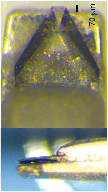
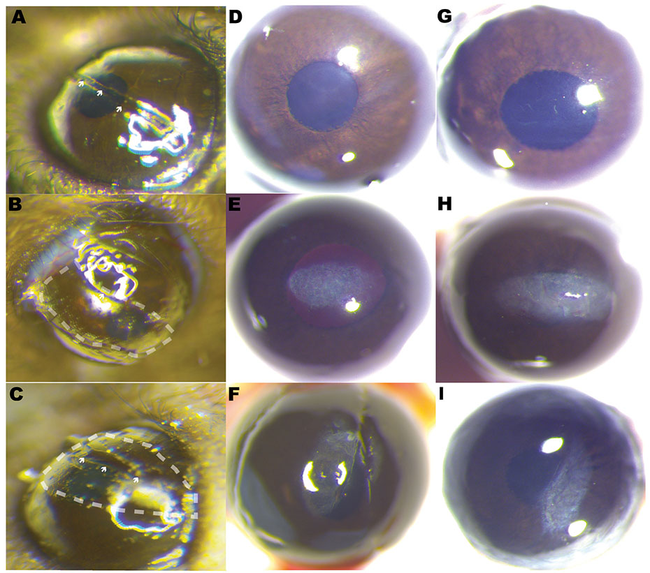
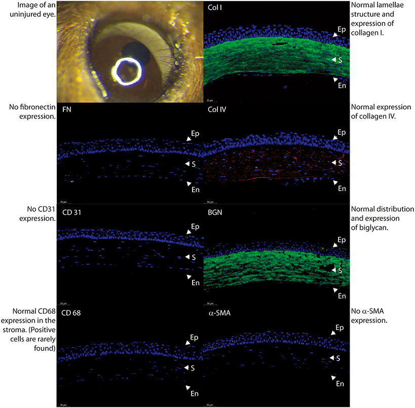
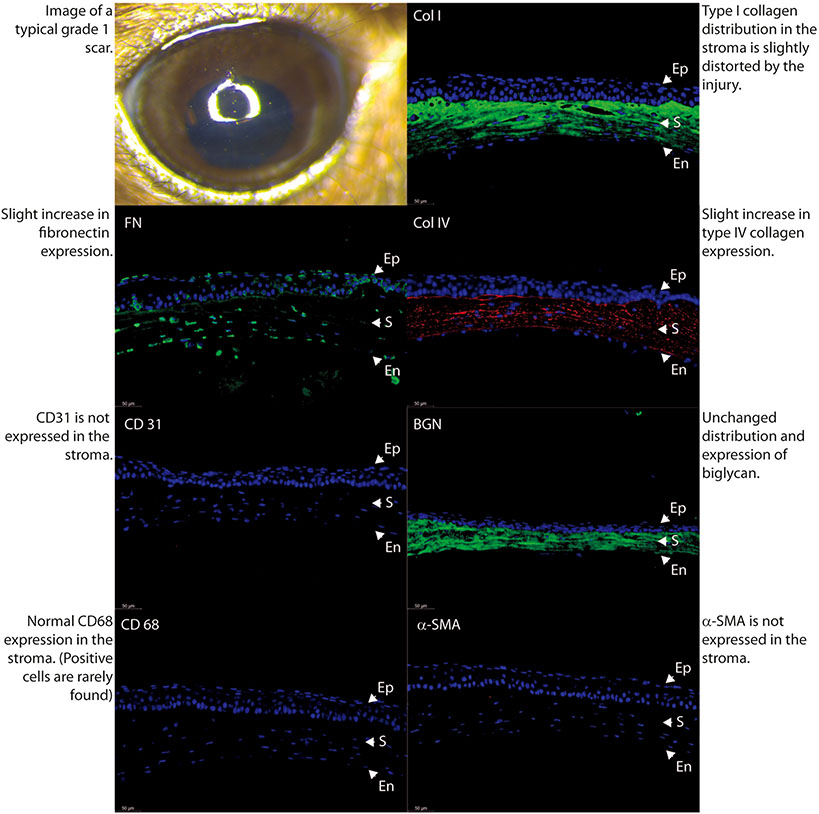
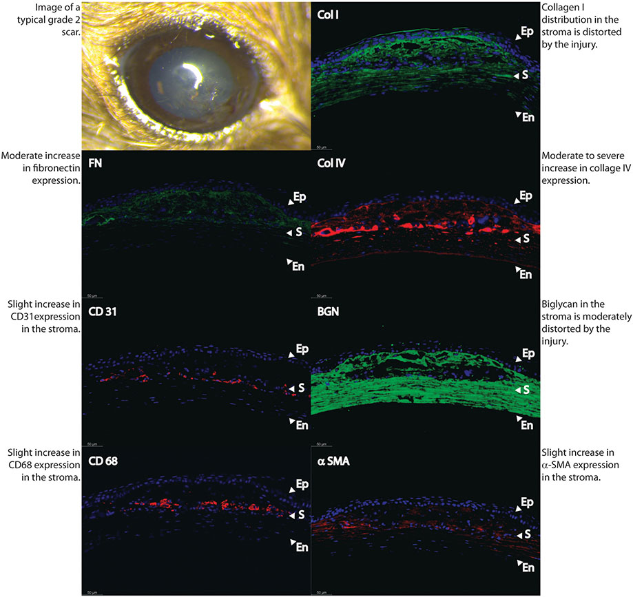
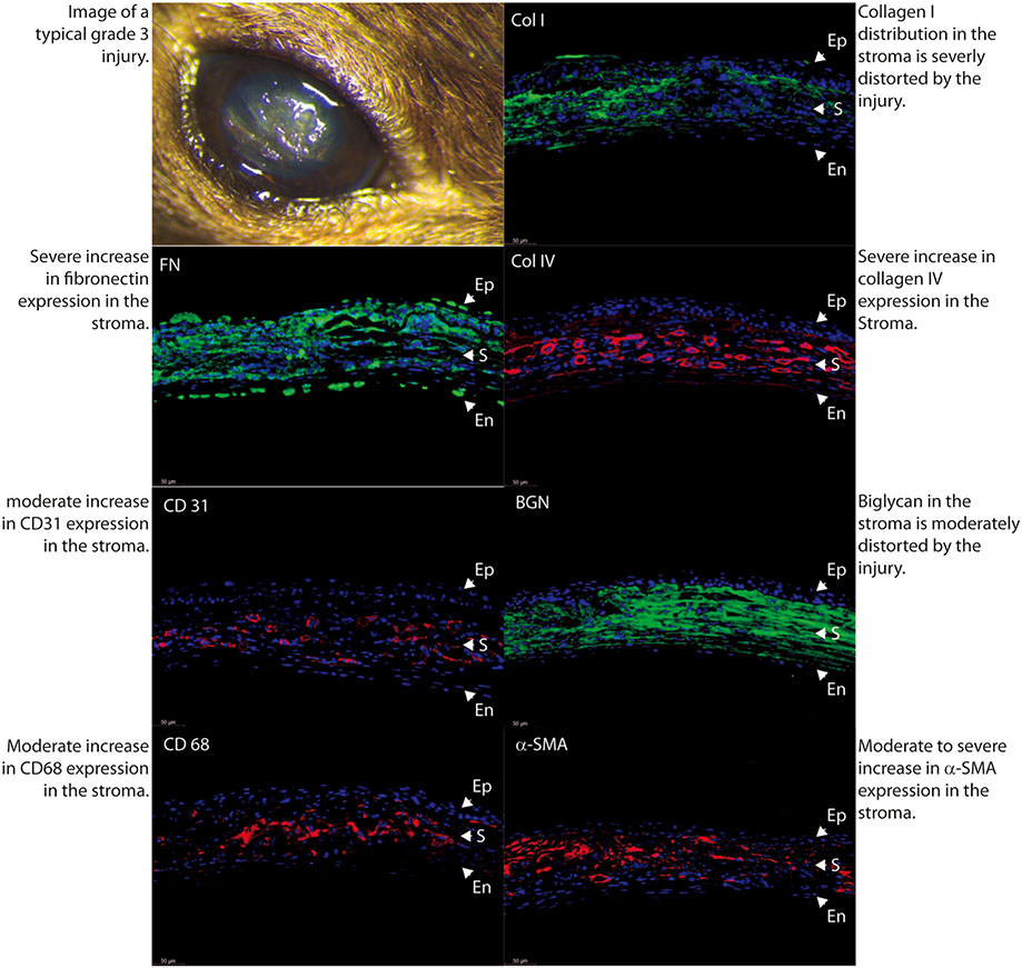
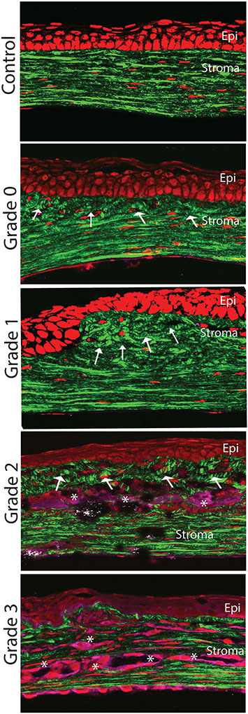
Materials and methods: We tested three stromal injury techniques in adult C57BL/6 mice. Technique 1, a lineal partial thickness keratotomy in the horizontal axis. Technique 2, corneal epithelial and stromal debridement using a diamond burr in the horizontal axis, and technique 3, a combination of techniques 1 and 2. To assess intra-observer and inter-observer agreement between two examiners evaluating formed stromal scars, stereo microscopic photographs of anterior segment were scored by two masked examiners at around 1-month. Depending on the severity of opacification and the area of involvement, scars were classified on a scale from 0 to 3 based on a modified Fantes haze scale. Extracellular matrix composition as well as matrix organization, macrophage infiltration and neovascularization were evaluated with immunofluorescence and second harmonic generation (SHG) microscopy.
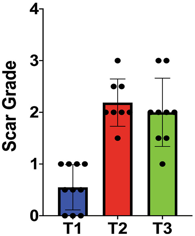
Results: Technique 1 created mild scars, with a score of 0.5 ± 0.43, while techniques 2 (score 2.1 ± 0.45) and 3 (score 2 ± 0.66), created dense scars with a higher score. A significant difference in scar severity score was noted between the 3 techniques (one way ANOVA, p < 0.0001). Masked graders demonstrated excellent agreement (intraclass correlation = 0.927 [95% confidence interval: 0.87-0.96]). The severity of scars noted at stereo microscopy correlated with the severity of changes in extracellular matrix in the stroma as demonstrated by the expression of collagens I, IV and fibronectin and evaluation of matrix hierarchical organization. In contrast to mild scarring, moderate and severe scars had increased expression of CD31 and CD68, markers of vascular endothelial cells and macrophages, respectively.
Conclusion: Mouse models of stromal scarring using simple surgical techniques are described. Corneal scars can be consistently classified by two observers. Grading of scar severity positively correlates with changes in extracellular matrix composition, disorganization and cell infiltration.
Keywords: Fibroblasts; Macrophages; Scar; Stroma; Wound.
Copyright © 2020 Elsevier Inc. All rights reserved.
Conflict of interest statement
Declaration of competing interest
None of the authors in this manuscript has any conflict of interest to disclose.
Figures
References
-
- Edelhauser HF. The balance between corneal transparency and edema: the Proctor Lecture. Invest Ophthalmol Vis Sci 2006;47:1754–67. - PubMed
Publication types
MeSH terms
Grants and funding
LinkOut - more resources
Full Text Sources
Other Literature Sources
Medical
