. 2020 Nov 29;21(23):9082.
doi: 10.3390/ijms21239082.

Gold Nanopeanuts as Prospective Support for Cisplatin in Glioblastoma Nano-Chemo-Radiotherapy

Affiliations

Gold Nanopeanuts as Prospective Support for Cisplatin in Glioblastoma Nano-Chemo-Radiotherapy

Joanna Depciuch et al. Int J Mol Sci. .

Abstract

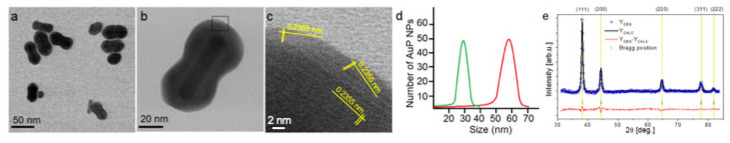
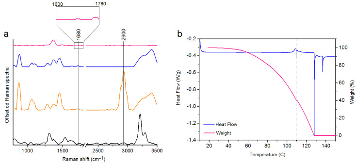
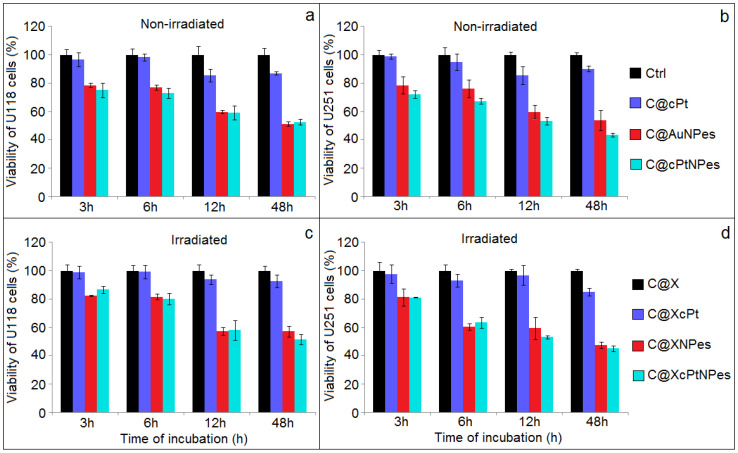
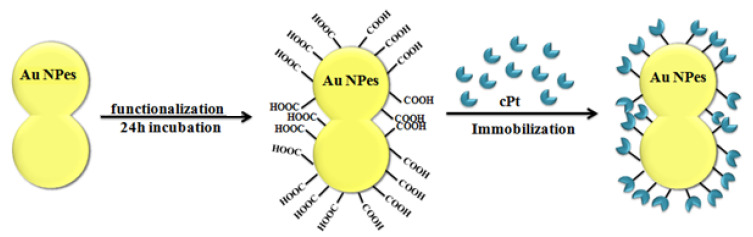
Herein, we propose newly designed and synthesized gold nanopeanuts (Au NPes) as supports for cisplatin (cPt) immobilization, dedicated to combined glioblastoma nano-chemo-radiotherapy. Au NPes offer a large active surface, which can be used for drugs immobilization. Transmission electron microscopy (TEM) revealed that the size of the synthesized Au NPes along the longitudinal axis is ~60 nm, while along the transverse axis ~20 nm. Raman, thermogravimetric analysis (TGA) and differential scanning calorimetry (DCS) measurements showed, that the created nanosystem is stable up to a temperature of 110 °C. MTT assay revealed, that the highest cell mortality was observed for cell lines subjected to nano-chemo-radiotherapy (20-55%). Hence, Au NPes with immobilized cPt (cPt@AuNPes) are a promising nanosystem to improve the therapeutic efficiency of combined nano-chemo-radiotherapy.

Keywords: biomedical application; gold nanopeanuts; new shape; support.


Conflict of interest statement

The authors declare no conflict of interest.

Figures

Figure 1
Figure 1
Overview BF STEM (a,b); HRSTEM image with indicated lattice distances of Au corresponding to the {111} planes. (c), size distribution of longitudinal (red curve) and transverse (green curve) axis (d) and XRD (e) taken for Au NPes. Experimental (blue dots) and calculated (solid black line) Rietveld refinement plot of the XRD pattern for the Au NPes. The red line (YOBS—YCALC) represents the difference between the observed and calculated data. Tick marks show allowed Bragg reflections (color online).
Figure 2
Figure 2
Raman spectra of: cPt (spectrum black), the pure MHDA (spectrum orange), the Au NPes + MHDA (spectrum blue), cPt@Au NPes (spectrum pink) (a); TGA and DSC data of cPt@Au NPes showing the solvent removal and decomposition of the product (b).
Figure 3
Figure 3
Comparison of the viability of U118 MG and U251 MG cell lines determined via the MTT assays in not-irradiated (a,b) and irradiated (c,d) cells by 2 Gy of X-rays after different time of incubation (3, 6, 24 and 48 h) with cisplatin, Au NPes and cPt@Au NPes and compared with controls.
Figure 4
Figure 4
Scheme of the Au NPes functionalization process.

References

    1. Glaser T., Han I., Wu L., Zeng X. Targeted nanotechnology in glioblastoma multiforme. Front. Pharmacol. 2017;8:166. doi: 10.3389/fphar.2017.00166. - DOI - PMC - PubMed
    1. Zakrzewska K.E., Samluk A., Wierzbicki M., Jaworski S., Kuwin M., Sawosz E., Chwalibog A., Pijanowska D.G., Pluta K.D. Analysis of the cytotoxicity of carbon-based nanoparticles, diamond and graphite, in human glioblastoma and hepatoma cell lines. PLoS ONE. 2015;10:e0122579. doi: 10.1371/journal.pone.0122579. - DOI - PMC - PubMed
    1. Holland E.C. Glioblastoma multiforme: The terminator. Proc. Natl. Acad. Sci. USA. 2000;97:6242–6244. doi: 10.1073/pnas.97.12.6242. - DOI - PMC - PubMed
    1. Rosa S., Connolly C., Schettino G., Butterworth K.T., Prise K.M. Biological mechanisms of gold nanoparticles radiosensitization. Cancer Nanotechnol. 2017;8:2. doi: 10.1186/s12645-017-0026-0. - DOI - PMC - PubMed
    1. Zhao X., Pan J., Li W., Yang W., Qin L., Pan Y. Gold nanoparticles enhance cisplatin delivery and potentiate chemotherapy by decompressing colorectal cancer vessels. Int. J. Nanomed. 2018;13:6207–6221. doi: 10.2147/IJN.S176928. - DOI - PMC - PubMed